
Evaluating the effectiveness of moxidectin treatment in mite-infested mice and development of an in-house PCR assay for detecting Myobia musculi DNA.
by M Portis, AD Floyd, CJ Perez, PS Huskey, DA Weiss, LG Coghlan, & F Benavides*
Department of Epigenetics and Molecular Carcinogenesis, The University of Texas MD Anderson Cancer Center, Smithville, Texas and The University of Texas Graduate School of Biomedical Sciences at Houston, Texas, USA.
 Correspondence: Dr. Fernando Benavides, Professor, Science Park, 1808 Park Road 1C -
P.O. Box 389, Smithville, Texas 78957, USA.
Tel: +1 512 2379343
Fax: +1 512 2372437
Email: fbenavid@mdanderson.org
Summary
Detection and eradication of fur mite infestations in mouse colonies can present a challenge to a laboratory animal facility. In 2011, an outbreak of Myobia musculi occurred in the University of Texas M.D. Anderson Cancer Center, Science Park animal facility, and the current case study describes the treatment with moxidectin, its effectiveness in controlling the outbreak, and the subsequent development of a PCR-based detection protocol. Methods of DNA collection using sterile swabs and fecal pellets were also evaluated. Results from the first mite check after the treatment indicated that moxidectin was highly effective in killing mites, but evidence of the infestation (mite body parts and eggs) was clearly visible throughout the duration of the monitoring period. Results from the PCR assay on swab samples were directly correlated with those of the conventional hair pluck samples, indicating that swabbing could be added to routine QA hair pluck monitoring procedures to increase the chances of detecting mites in laboratory mice. However, it is essential to keep in mind that positive PCR results may not necessarily indicate an active infestation but only the presence of mite DNA in the hair coats of mice.
Introduction
The presence of ectoparasites in research facilities has been shown to induce homeostatic stress on animals, prompting behavioral, physiological or immunological responses that can potentially alter research results (Morita et al., 1999; Pocanke et al.) .
Several species of fur mites occur in laboratory rodents, including Myobia musculi, Myocoptes musculinus, and Radfordia affinis. M. musculi is a non-burrowing fur mite found primarily on the head and neck region. This species thrives in the epidermal environment, completing its entire 23 day life cycle on a single host by deriving nourishment from sebaceous secretions and lymph produced by mite-associated ulcerative dermatitis that often accompanies mite infestation (Weisbroth et al., 1974; Friedman & Weisbroth, 1975). Adult females are capable of producing fertilized eggs within 24 hours of reaching maturity, and the eggs hatch in approximately seven days. Most species of fur mites are primarily transmitted by direct contact with an infested host, and transfer to neonate mice can occur as early as seven days of age, around the time the fur coat develops. M. musculi has been documented to survive on deceased hosts for up to four days and is the most clinically significant mite found in mice (Percy, 2007).
Detection methods for fur mites include the cellophane tape-test, hair pluck test, skin scraping analysis, stereomicroscopic pelt examination, and polymerase chain reaction (PCR) assay (Rice et al., 2013). Accuracy of results from different detection methods can vary based on the intensity of the mite infestation and the age, strain, housing conditions and hair cycle stage of afflicted mice (Jensen et al., 2013; Lindstrom et al., 2011; Metcalf Pate et al., 2011; Weiss et al., 2012; Karlsson et al., 2014) . Effectiveness of mite detection strategies also varies among the sampling types: a few of the tests can be conducted postmortem (which is not always desirable), but the most widely used methods, hair plucking, tape test and PCR, can be performed on live animals (Metcalf Pate et al., 2011) .
Mice experiencing light acariasis may exhibit no apparent clinical symptoms of infestation, but common pathological effects associated with heavier ectoparasite loads range from mild inflammation to serious epidermal lesions, including pruritus, ulceration and pyoderma (Morita et al., 1999; Rice et al., 2013) . Secondary symptoms can occur, and include wasting syndrome, alopecia, self-inflicted excoriation and infection of abrasions. Immunological sequelae related to M. musculi infestation have also been observed, including hypoalbuminemia, lymphoadenopathy, hypergammaglobulinemia and an increase in mast cell infiltration of epidermal lesions. Additionally, intensity of the pathological effects of murine acariasis can vary depending on mouse strain (Johnston et al., 2009) .
Eradication of fur mite infestations in mouse colonies can present serious challenges. Previous studies have demonstrated the efficacy of ivermectin-based compounds in controlling fur mite outbreaks (Papini & Marconcini, 1991). Ivermectin is a commonly used broad spectrum anti-parasitic medication that can be applied directly to the skin or introduced into the diet for treatment purposes (Ricart Arbona et al., 2010a, 2001b) . Ivermectin has been used in laboratory animals with some success, but because little is known about the pharmacokinetic properties in mice, neurotoxicity as a potential side effect may be a concern in some mouse strains (Kwei et al., 1999) . Though ivermectin compounds have been highly effective in short-term control of fur mites, reapplication is necessary to accommodate all stages of the life cycle, which can increase treatment cost and time, challenging the practicality of ivermectin use as a long term parasiticide. A promising alternative is moxidectin, a milbemycin compound chemically related to ivermectin with a similar mode of action (Pullium et al., 2005; Pollicino et al., 2008) . This compound is widely used as a safe topical pour-on acaracide in cattle and other livestock (Cydectin, Fort Dodge), though its use in laboratory rodents has been limited.
In 2011, an outbreak of M. musculi occurred in the University of Texas M.D. Anderson Cancer Center, Science Park animal facility in Smithville Texas. The infestation was initially detected through routine hair pluck samples from sentinel animals. Examination of multiple additional samples from selected cages confirmed the presence of live M. musculi mites in two separate rooms. Consequently, the decision was made to treat with moxidectin, because it is safe, inexpensive and readily available. During the treatment and monitoring process, we sought to develop an in-house PCR assay to detect the presence of M. musculi and to apply a treatment regimen that would effectively eradicate the infestation, while simultaneously employing a monitoring strategy to detect any re-infestation that occurred. The current case study describes the treatment regimen that was implemented, its effectiveness in controlling or eliminating the outbreak, and the development of an in-house PCR test to accompany traditional hair pluck monitoring to detect M. musculi infestations.
Materials and methods
Animal Housing and Maintenance
All of the mice were housed in an AAALAC-accredited facility in Individually Ventilated Cages (IVC), (Micro-isolator Housing Unit; Lab Products, Maywood, NJ) sterilized together with aspen chip bedding (Aspen Sani-chips; P.J. Murphy Forest Products Corporaton, Montville, NJ); the bedding and the cages were changed every 9 days in a mobile cage changing station (NuAire, Plymouth, MN) which provides ISO Class 4 protection. Wayne 22/5 Rodent Diet #8640 (Harlan Teklad, Madison, WI) was provided ad libitum and acidified reverse osmosis water was provided by an automated system. Room SRG1.121 is approximately 884 sq. ft maintaining 12 IVC racks. Room temperature and humidity were 20 to 22° Celsius and 55% respectively, with a 14 hour light and 10 hour dark cycle and a minimum of 12 air changes per hour. Outbred Cr:SW (Swiss Webster) sentinel animals are maintained in each animal room and sentinel workups are performed semi-annually for rodents which includes serology and parasitology.
Moxidectin Treatment
Treatment interval and number of treatments were developed based on other reports in the literature (Pullium et al., 2005) and the M. musculi life cycle. Animals in rooms found to have active infestations were treated twice, 10 days apart. Using an automatic repeating pipet, 10 μl of a Cydectin (0.5% moxidectin; Fort Dodge)/corn oil mixture (50:50) was applied to the skin on the dorsum of each haired animal between the shoulder blades (1 mg/kg per 25 g mouse). Treatment dates correlated with regularly scheduled change out days when all caging components (cages, wires and lids) were replaced with newly cleaned components. Pups that did not have a hair coat during the first treatment date were subsequently treated on the second date.
Monitoring by Hair Plucking
Seven and a half weeks after the final treatment, a total of 34 cages from the larger of two treated rooms were selected for hair pluck monitoring based on their infestation history. Each mouse in the selected cages was restrained, and two or three small groups of hair were plucked from the dorsum of each animal just behind the head. These samples were laid flat on a standard microscope slide and a few drops of mineral oil and a coverslip were applied. Prepared slides were examined under a microscope (4x, 0.10 scanning power) for any sign of mite infestation, which included the presence of live mites, eggs (both empty and containing embryos) or parts of dead mites. Any sign of mites was considered a positive result. Selected cages were continually monitored at regular intervals up to 42 weeks after the second treatment, and the number of empty egg casings, intact eggs and mites were recorded for each cage. To assist in the development of our mite PCR testing, fecal pellets were collected from animals in known positive cages, and known positive animals were swabbed with a sterile cotton tipped applicator.
Monitoring by PCR Analysis
During the monitoring period, intact mites, fecal pellets and swab samples were collected from selected cages. Genomic DNA was extracted from adult M. musculi (collected and identified under a dissection microscope) and from swab samples using a modified simplified extraction method (Laird et al., 1991). Fecal pellet DNA was extracted using the Qiagen Stool Kit (Qiagen, Inc., Valencia, CA). Primers for detecting M. musculi were designed based on the 18S rRNA gene (gb JF934703.1) from Feldman and Ntenda (Feldman & Ntenda, 2011) using Vector NTI software (Invitrogen Corp., Carlsbad, CA). The sequences are as follows: forward 5’ – AAGGATCTATTGGAGGGCAAGTCTC – 3’, reverse 5’ – GGCTGATCGCTGGTTGGCATA – 3’. Reaction mixtures contained 2 μM of each oligonucleotide primer at 1 μM, PCR buffer (10 mM Tris-HCl, 1.5 mM MgCl2, 50 mM KCl, 1.5 U of AmpliTaq DNA polymerase (Life Technologies, Grand Island, NY), and DNA template. All reactions were performed in a 25 μL volume in an Eppendorf Mastercycler Gradient® thermocycler under the following cycling conditions: 95oC, 1 minute; then 40 cycles of 95oC, 35 seconds; 60oC, 45 seconds; 68oC, 45 seconds; followed by a final incubation at 68oC for 10 minutes. Electrophoresis of the PCR product was carried out using 2% agarose gel, and the appearance of a band at 522 bp was considered a positive result. To positively identify the PCR product as that of M. musculi DNA, the amplicon was purified by silica columns (Qiagen Inc., Chatsworth, CA.) and sequenced with the Taq dideoxy-chain termination method (ABI PRISM® BigDye® Terminator ver. 3, Applied Biosystems Inc., Foster City, CA) in an ABI3130XL instrument (Life Technologies, Grand Island, NY). The sequence identity was confirmed with the BLASTn algorithm (Ver. 2.2.26+) on the NIH Genbank nr database.
Results
Two treatments 10 days apart were conducted in a large animal room containing approximately 3,660 mice in August 2011. Seven and a half weeks after the second treatment, 34 cages previously known to be positive (91 mice total) were selected (based on factors such as strain and permission from investigators) for continual collection of specimens to check for the status of mite infestation using the hair pluck method. Twelve of these cages were considered positive for mite infestation due to the presence of numerous empty egg casings, intact eggs, dead mites and other mite related debris in the initial hair pluck sample (Mite Check #1, Figure 1). No live mites were identified in any of the positive samples. At 10 weeks post treatment (Mite Check #2), nine of the 12 initially positive cages were remaining (25 mice total). Five cages were still positive, while four cages had negative hair pluck results, with no visible signs of mite infestation. Interestingly, at just under 15 weeks post treatment, hair plucks from all eight cages still available for monitoring (23 mice total) showed residual evidence of mites, ranging from a few mite parts to numerous empty and intact eggs, even though four of the cages were negative at the previous mite check. This anomaly was likely due to the inherent variability of the hair pluck procedure rather than re-infestation, as no live mites were observed in any samples collected after treatment. These cages all remained positive by hair pluck in subsequent mite checks, with only 5 cages (12 mice total) still available for monitoring at 21 weeks after treatment (Mite Check #5).
|
Figure 1. Time line of events during mite treatments and checking. Positive status was assigned to hair pluck samples when mites, intact eggs and/or empty eggs were present. PCR results were considered positive when a unique band appeared at 522 bp on agarose gel electrophoresis, the expected size of the specific amplification of the M. musculi 18S rRNA gene. Cells marked with an asterisk (*) indicate the time period in which the PCR detection methods were being developed, and no swab samples were collected at these times. PCR testing of swab samples began at 29 weeks post treatment.
Click image to enlarge |
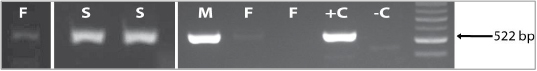 |
Figure 2. Detection of M. musculi DNA through conventional PCR methods. Both swab samples (S) produced strong positive bands at 522 bp. Two of five fecal samples (F) produced faint positive bands. These samples were compared to DNA isolated from whole M. musculi mites (M), known positive fecal pellets (+C) and a non-template control (-C). Breaks in the gel picture indicate areas where a number of negative samples were cut out to simplify the gel image for the figure. The bright band on the DNA ladder indicates 500 bp. Identity of the amplicon was confirmed by sequencing and alignment with M. musculi sequences stored in the NIH Genbank database. See Materials and Methods for details.
Click image to enlarge |
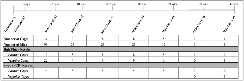
At 29 weeks post treatment (Mite Check #6), the mite PCR protocol was established and ready for testing, so swab samples were collected in addition to fecal samples and hair plucks to test the efficacy of the mite PCR analysis. The two remaining cages (5 mice) maintained their positive status via hair pluck and by PCR analysis using primers developed by our lab. Gel visualization of PCR product resulted in a clearly positive band at 522 bp for each swab sample (one per cage), and two of the five fecal DNA samples (one per mouse) produced a faint positive band (Figure 2). To further confirm our findings, the resulting bands were compared to the positive control extracted from whole M. musculi mites and a non-template control containing water. Swab samples resulted in better quality DNA at higher concentrations than that extracted from the fecal samples, possibly due to low amounts of DNA in fecal pellets, the presence of DNA inhibitors in fecal matter, and/or improved collection of mite and egg matter and debris by swabbing the whole mouse body.
In order to verify the identity of the resulting bands as M. musculi DNA, the PCR product was purified, quantified and sent to the Science Park Molecular Biology Core for sequencing. Identity of the nucleotide sequence of the amplified 18S rRNA gene fragment was confirmed by alignment with sequences stored in the NIH Genbank database. Our PCR product matched the M. musculi 18S ribosomal RNA gene at 99 percent certainty (Accession # JF934703.1 and JF83489.5).
Discussion
Overall, the treatment was highly successful in quickly getting the outbreak under control. Mice responded well to treatments, and there were no observed adverse effects on the various mouse strains housed in the treated rooms, including several inbred strains (C57BL/6, FVB/N, 129S6, and mixed backgrounds), transgenic, knock-out and knock-in animals. Based on visual observations in hair pluck samples, the application of moxidectin killed live mites, but apparently not embryonic mites or eggs. Consequently, we believe that viable eggs continued to incubate, hatched, and newly emerged nymphs were killed during the second scheduled treatment. Mite DNA (and, consequently, a positive PCR result) was isolated from samples collected from mice harboring residual dead mites, mite parts and eggs (as indicated by hair pluck), so it is imperative to consider this when interpreting results, as there may not be an active infestation or live mites although mite DNA is present.
We successfully developed an in-house PCR assay and identified an efficient collection method to test for the presence of M. musculi. The results of the fur swab PCR were directly correlated to corresponding hair pluck results. Positive bands were clearly visible at the expected size of 522 bp and a match to M. musculi DNA was confirmed through sequencing of the PCR product. The swabbing technique was simple, efficient and reliable, and DNA collected in this manner produced clear PCR amplicons that could be easily interpreted from gel visualization. Fecal pellets were also fairly easy to obtain, but were not the most reliable sampling technique, as they yielded only faintly positive results for the presence of mites. With the successful development of a PCR mite test, current QA procedures for detecting mites could be amended to add swabbing as a supplemental method of mite detection.
In summary, moxidectin treatment of a mite infested laboratory mouse colony was successful in quickly controlling a M. musculi outbreak. Additional cleaning and decontamination procedures, such as washing racks, caging components and surfaces in the room, is highly recommended to further protect against re-infestation by “renegade” mites hiding in cracks and crevasses of equipment. Routine hair pluck procedures remain an important method for mite detection, and this outbreak contributed to the decision to increase our sentinel QA monitoring schedule from two to four times per year for earlier detection of future mite infestations. This monitoring can be supported by hair coat swabbing for PCR testing, which can provide higher sensitivity for detecting mite infestations at an earlier stage. However, it is not recommended to utilize only the hair swabbing-PCR procedure in the follow-up after treatment, as positive PCR results may not necessarily indicate an active infestation and may simply occur from the presence of residual mite DNA in the hair coats of laboratory mice, even months after the eradication.
Acknowledgements
We thank the Molecular Biology Facility Core for DNA sequencing. This work was supported by the NIH/NCI under award number P30CA016672 and used the CCSG RASF-S Animal Resource and Genetic Services.
References
- Feldman SH & AM Ntenda: Phylogenetic analysis of Myobia musculi (Schranck, 1781) by using the 18S small ribosomal subunit sequence. Comp. Med. 2011, 61, 484-491.
- Friedman S & SH Weisbroth: The parasitic ecology of the rodent mite Myobia musculi. II. Genetic factors. Lab. Anim. Sci. 1975, 25, 440-445.
- Jensen, ES, KP Allen, KS Henderson, A Szabo & JD Thulin: PCR testing of a ventilated caging system to detect murine fur mites. J. Am. Assoc. Lab. Anim. Sci. 2013, 52, 28-33.
- Johnston NA, RA Trammell, S Ball-Kell, S Verhulst & LA Toth: Assessment of immune activation in mice before and after eradication of mite infestation. J. Am. Assoc. Lab. Anim. Sci. 2009, 48, 371-377.
- Karlsson, EM, LM Pearson, KM Kuzma & TH Burkholder: Combined evaluation of commonly used techniques, including PCR, for diagnosis of mouse fur mites. J. Am. Assoc. Lab. Anim. Sci. 2014, 53, 69-73.
- Kwei GY, RF Alvaro, Q Chen, HJ Jenkins, CE Hop, CA Keohane, VT Ly, JR Strauss, RW Wang, Z Wang, TR Pippert & DR Umbenhauer: Disposition of ivermectin and cyclosporin A in CF-1 mice deficient in mdr1a P-glycoprotein. Drug Metab. Dispos. 1999, 27, 581-587.
- Laird PW, A Zijderveld, K Linders, MA Rudnicki, R Jaenisch & A Berns: Simplified mammalian DNA isolation procedure. Nucleic Acids Res. 1991, 19, 4293.
- Lindstrom KE, LG Carbone, DE Kellar, MS Mayorga & JD Wilkerson: Soiled bedding sentinels for the detection of fur mites in mice. J. Am. Assoc. Lab. Anim. Sci. 2011, 50, 54-60.
- Metcalf Pate KA, KA Rice, R Wrighten & J Watson: Effect of sampling strategy on the detection of fur mites within a naturally infested colony of mice (Mus musculus). J. Am. Assoc. Lab. Anim. Sci. 2011, 50, 337-343.
- Morita E, S Kaneko, T Hiragun, H Shindo, T Tanaka, T Furukawa, A Nobukiyo & S Yamamoto: Fur mites induce dermatitis associated with IgE hyperproduction in an inbred strain of mice, NC/Kuj. J. Dermatol. Sci. 1999, 19, 37-43.
- Papini R & A Marconcini: Treatment with Ivermectin in drinking water against Myobia musculi and Myocoptes musculinus mange in naturally infected laboratory mice. Angew. Parasitol. 1991, 32, 11-13.
- Percy DH & SW. Barthold: Pathology of laboratory rodents and rabbits, 3rd Edition. Wiley–Blackwell, 2007, pp 3–124.
- Pochanke V, S Hatak, H Hengartner, RM Zinkernagel & KD McCoy: Induction of IgE and allergic-type responses in fur mite-infested mice. Eur. J. Immunol. 2006, 36, 2434-2445.
- Pollicino, P, L Rossi, L Rambozzi, AM Farca & A Peano: 2008. Oral administration of moxidectin for treatment of murine acariosis due to Radfordia affinis. Vet. Parasitol. 2008, 151, 355-357.
- Pullium JK, WI Brooks, AD Langley & MJ Huerkamp: A single dose of topical moxidectin as an effective treatment for murine acariasis due to Myocoptes musculinus. Contemp. Top. Lab. Anim. Sci.2005, 44, 26-28.
- Ricart Arbona RJ, NS Lipman, ER Riedel, & FR Wolf:. Treatment and eradication of murine fur mites: I. Toxicologic evaluation of ivermectin-compounded feed. J. Am. Assoc. Lab. Anim. Sci.2010a, 49, 564-570.
- Ricart Arbona RJ, NS Lipman & FR Wolf:. Treatment and eradication of murine fur mites: III. Treatment of a large mouse colony with ivermectin-compounded feed. J. Am. Assoc. Lab. Anim. Sci.2010b 49, 633-637.
- Rice KA, LK Albacarys, KA Metcalf Pate, C Perkins, KS Henderson & J Watson. Evaluation of Diagnostic Methods for Myocoptes musculinus According to Age and Treatment Status of Mice (Mus musculus). J. Am. Assoc. Lab. Anim. Sci. 2013, 52, 773-781.
- Weisbroth SH, S Friedman, M Powell & S Scher: The parasitic ecology of the rodent mite Myobia musculi. I. Grooming factors. Lab Anim Sci. 1974, 24, 510-516.
- Weiss EE, KD Evans & SM Griffey: Comparison of a fur mite PCR assay and the tape test for initial and posttreatment diagnosis during a natural infection. J. Am. Assoc. Lab. Anim. Sci. 2012, 51, 574-578.