
An in vivo permeability test protocol using iohexol to reduce and refine the use of laboratory rats in intestinal damage assessment
by Rafael Frias1*, Arthur C. Ouwehand2,3, Ulla-Marjut Jaakkola1, Thomas Spillmann4, and Miguel Gueimonde2,5
1Central Animal Laboratory, University of Turku, Kiinanmyllynkatu
10, C6, FIN-20520, Turku, Finland;
2Functional Foods Forum, University of Turku, Finland;
3Health and Nutrition, Danisco, Kantvik, Finland;
4Dept. of Equine and Small Animal Medicine, Faculty of
Veterinary Medicine, University of Helsinki, Finland;
5Instituto de Productos Lácteos de Asturias (IPLA),
Consejo Superior de Investigaciones Científicas (CSIC),
Villaviciosa, Asturias, Spain.
 Correspondence: Rafael Frias
Central Animal Laboratory, University of Turku,
Kiinanmyllynkatu 10, C6, FIN-20520, Turku, Finland
Tel +358 2333 7625
Fax +358 2333 7343
Email raffri@utu.fi
Summary
Assessment of intestinal damage in laboratory rats with experimentally-induced enteropathies is usually carried out by collecting and morphological interpreting tissue samples obtained surgically, endoscopically or at necropsy. Alternatively, changes in the gut mucosa may be less invasively evaluated with intestinal permeability (IP) tests. In contrast to human and veterinary patients, IP test protocols in laboratory rats have been highly variable, which may account for the limited use of this approach by investigators when evaluating intestinal damage. The objective of this study was to establish a refined IP test protocol using iohexol in rats that is able to differentiate between healthy rats and individuals with enteropathies. Iohexol was administered by oral gavage to twenty-eight Sprague-Dawley rats, before and after the induction of inflammatory bowel disease (IBD) with dextran sulphate sodium (DSS). Urine was cumulatively recovered during 24 h, and the presence of iohexol was measured by high-performance liquid chromatography with ultraviolet detection. The median percentage (and interquartile range) of administered iohexol in urine of healthy rats was 0.54% (0.36–0.75%), whereas the respective value in rats with enteropathies was 11.42% (5.58–15.37%). The significant difference (P < 0.001) in the urinary recovery of iohexol demonstrated sufficient sensitivity of the test protocol to clearly discriminate between healthy and affected rats.
Introduction
The degree of damage to the intestinal mucosa of laboratory animals is commonly evaluated in research projects aimed at the diagnosis and follow-up of intestinal integrity, especially in studies that involve experimentally-induced gastrointestinal diseases or in the development of potential therapeutic agents. The assessment of intestinal mucosal integrity is typically based on the interpretation of tissue specimens collected surgically, endoscopically or at necropsy. However, this approach has shortcomings from scientific and animal welfare perspectives. Histological samples may be incorrectly interpreted, and they only provide morphological insights rather than information on intestinal wall permeability. Furthermore, an intestinal biopsy is an invasive procedure requiring anaesthesia, and additional animals serving as controls must be used (Kim & Berstad, 1992; Hall, 1994; Ahn et al., 2001; Hoffmann et al., 2002; Jurjus et al., 2004; Chen et al. 2007). IP tests, however, allow the non-invasive assessment of intestinal mucosal integrity. These tests have been successfully applied in humans and a variety of animal species to detect primary intestinal permeability defects, and also to monitor recovery from intestinal damage after therapy in both clinical and research settings (Bjarnason et al., 1995; Hollander, 1999; Hall, 1999).
IP test protocols have frequently involved the use of 51chromium-labelled ethylenediamine tetra-acetic acid (51Cr-EDTA) and a variety of sugars (e.g. lactulose and rhamnose) as probe markers. However, disadvantages associated with the use of 51Cr-EDTA radioactivity and bacterial degradation of the saccharides has led to the search for better probe candidates. More recently, iohexol, an iodinated contrast agent commonly used in medical imaging, has also been successfully applied as an IP marker for the non-invasive screening of intestinal damage in laboratory rats and humans. The main advantages of using this substance are that it is non-radioactive, biologically inert, widely available in radiological units, relatively inexpensive, and also allows the simultaneous examination of the gastrointestinal tract using other imaging techniques (Stordahl 1988; Bjarnason et al., 1995; Halme et al., 1993; Halme et al., 1997; Halme et al., 2000; Hall, 1999; Andersen et al., 2001; Frias et al., 2009).
Furthermore, IP testing methodology has essentially been standardized in human and veterinary patients, although in laboratory rats there is a substantial lack of uniformity in testing protocols. For example, IP testing has been inconsistently attempted in this species in vivo on either anaesthetized or conscious subjects, after notably variable timed urinary recoveries, from tissue specimens collected from anaesthetized animals via invasive methods, or ex vivo during post-mortem examinations (Bjarnason et al., 1985; Willoughby et al, 1996; Andersen et al., 2001; Milde et al., 2003).
The objective of this study was to establish an improved IP test protocol using iohexol that is able to discriminate between healthy and affected rats, and that is consistent with Russell and Burch’s guiding principles on the reduction and refinement of laboratory animal use.
Materials and methods
Experimental animals, housing and husbandry
Thirty female adult Hsd:Sprague Dawley®™SD®™ (SD) rats obtained from a
breeding colony kept under semi-barrier conditions at the Central
Animal Laboratory, University of Turku, Finland, were used in this
study, which was part of another study reported previously (Frias et al., 2009). At the commencement of the study, the rats were 12 weeks old and
ranged in body weight from 200 to 250 g. The rats were housed in
groups of six, and grouping was decided based on rat availability from
the breeding colony. They were maintained in stainless steel cages
(59.5 x 38.0 x 20.0 cm) with solid bottoms and Aspen chips as bedding
(Tapvei Ltd, Kaavi, Finland), with enrichment consisting of an Iglo
and some nesting material. Cage change was undertaken twice a week.
The environment in the room consisted of a temperature range of 20 to
23 °C, a relative humidity of 50 to 60%, and artificial illumination
with a 12-h light/dark cycle (lights on at 06:00 am). Throughout the
study period, all the rats were fed a standard rat chow (SDS, Special
Diet Services, Whitham, Essex, UK) ad libitum, and tap water
was provided without restrictions in polycarbonate bottles. Prior to
the exposure to dextran sulphate sodium (DSS), all the rats were
acclimatized for 21 days and were determined to be healthy on the
basis of individual physical examinations, and pathogen-free based on
the results of routine microbiological screening performed on the
colony in accordance with European recommendations (Nicklas et al., 2002).
Ethical statement
The rats were cared for and used in accordance with Finnish
legislation and Council of Europe Convention ETS 123 on the use of
vertebrate animals for scientific purposes (Council of Europe, 1986; Finnish Government, 1985; Finnish
Government, 1996), and the experimental protocol was part of a project approved by
the Ethics Committee for Animal Experiments of the University of
Turku, Finland.
Study design
The oral iohexol IP test in urine was carried out twice in each of the
thirty SD rats included in this study, once before and then seven days
after the experimental induction of gastrointestinal damage.
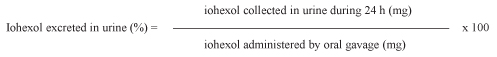
Table 1. Formula to calculate the percentage recovery of orally ingested iohexol in rat urine.
Induction of gastrointestinal damage
Gastrointestinal damage was induced by the seven-day administration of
5% dextran sulphate sodium (DSS) in drinking water, which has been
shown to produce symptoms in laboratory rats comparable to the
inflammatory bowel disease (IBD) observed in humans (Gaudio et al., 1999; Chen et al., 2007; Frias et al., 2009).
Oral iohexol IP test measured in urine
Immediately before the test was carried out, the body weight of the
rats was measured. Next, 1 ml of Omnipaque 300® (iohexol, 647.1 mg/mL)
was dosed intragastrically to each rat using a feeding tube. No
sedative drug was used before, during or after administration. The
animals were placed in individual metabolic cages for 24 h for urine
collection. After all urine had been recovered, the volumes were
recorded and the samples frozen at -18 ºC until later analysis. If
oesophageal reflux of iohexol or faecal contamination of urine was
observed, the test was cancelled.
Laboratory analysis of iohexol and creatinine
Iohexol concentration in urine was analysed by high-performance liquid
chromatography with ultraviolet detection [(HPLC)-UV] after solid
phase extraction according to a previously published method (Klenner et al., 2007). The formula to calculate the percentage of iohexol excreted in
urine is provided in Table 1.
To assess the possible toxic effects of DSS on kidney function, creatinine was determined in all urine samples using a Konelab 30i automatic analyser (Thermo Scientific, Waltham, MA, USA). The iohexol-to-creatinine ratio was calculated similarly to the urinary protein-to-creatinine ratio for the assessment of proteinuria in dogs (White et al., 1984; Grauer et al., 1985).
Statistical methods
Statistical analysis was performed using SPSS 11.0 software (SPSS Inc,
Chicago, IL, USA). The data were analysed with the Wilcoxon
signed-ranks test, and were expressed as the median and interquartile
range (IQR).
Results
Twenty-eight SD rats were enrolled in the study after the exclusion of two because of oesophageal reflux. Rats exposed to DSS showed evidence of ulcerative colitis based on physical examination and evidence of changes in faecal consistency, diarrhoea and haematochezia. (Gaudio et al., 1999) All serum creatinine concentrations (n = 27) suggested normal renal function.
The median (IQR) percentage (%) of administered iohexol in urine of healthy rats was 0.54% (0.36–0.75%), whereas the respective value after DSS administration was 11.42% (5.58–15.37%). The median (IQR) iohexol/creatinine ratio was 0.05 (0.03–0.06) in healthy rats and 1.38 (0.76–2.49) in rats with IBD.
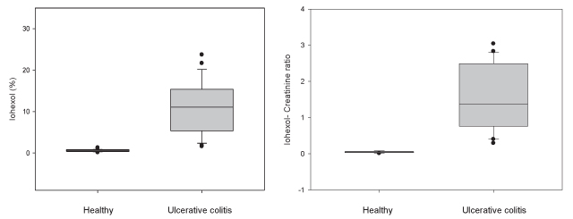
Figure 1 presents percentile plots of urinary iohexol and the iohexol-creatinine ratio before and after the induction of ulcerative colitis by adding 5% DSS to the drinking water for seven days. Nonparametric comparison of the urinary excretion of iohexol as well as the iohexol/creatinine ratio demonstrated a statistically significant difference (P < 0.001) between healthy rats and those with colitis.
|
Figure 1. Statistical analysis of the relationships between the levels of iohexol and the iohexol-creatinine ratio before and after induction of ulcerative colitis (n = 28). The line in the box represents the median (50%); the lower line represents the lower (25%) quartile, and the upper line represents the upper (75%) quartile. The limits of the upper and lower vertical lines indicate the highest and lowest data values, respectively. The separate asterisks indicate outliers.
Click image to enlarge |
Table 2. Individual results from the iohexol intestinal permeability test before and after administration of DSS for induction of intestinal damage in laboratory SD rats.
| Iohexol | Iohexol/creatinine ratio | |||
| Rat # | Before | After | Before | After |
| 1 | 0,83 | 9,74 | 0,07 | 0,72 |
| 2 | 1,15 | 5,58 | 0,08 | 1,46 |
| 3 | 0,65 | 3,20 | 0,05 | 0,76 |
| 4 | 0,19 | 5,25 | 0,02 | 0,88 |
| 5 | 0,75 | 2,42 | 0,06 | 0,4 |
| 6 | 0,75 | 1,84 | 0,05 | 0,29 |
| 7 | 0,59 | 12,54 | 0,05 | 0,50 |
| 8 | 0,12 | 20,02 | 0,01 | 2,55 |
| 9 | 0,36 | 9,82 | 0,04 | 2,77 |
| 10 | 0,43 | 17,2 | 0,04 | 2,49 |
| 11 | 0,85 | 18,77 | 0,08 | 2,83 |
| 12 | 0,81 | 1,57 | 0,07 | 0,48 |
| 13 | 1,25 | 3,94 | n.d. | n.d. |
| 14 | 0,85 | 15,36 | 0,08 | 2,3 |
| 15 | 0,52 | 10,7 | 0,05 | 2,43 |
| 16 | 0,44 | 4,16 | 0,03 | 0,41 |
| 17 | 0,54 | 15,83 | 0,06 | 1,61 |
| 18 | 0,63 | 23,71 | 0,05 | 2,79 |
| 19 | 0,31 | 8,77 | 0,03 | 2,67 |
| 20 | 0,32 | 8,77 | 0,02 | 1,33 |
| 21 | 0,54 | 12,05 | 0,04 | 1,34 |
| 22 | 0,37 | 21,64 | 0,03 | 3,04 |
| 23 | 0,48 | 10,13 | 0,04 | 1,51 |
| 24 | 0,30 | 15,37 | 0,03 | 1,51 |
| 25 | 0,30 | 12,05 | 0,03 | 1,24 |
| 26 | 0,56 | 12 | 0,05 | 1,17 |
| 27 | 0,81 | 12,58 | 0,06 | 1,38 |
| 28 | 0,38 | 11,42 | 0,03 | 0,96 |
* Not determined, n.d.
Discussion
IP may be assessed by measuring the cumulative urinary excretion of an orally-administered dose of iohexol. An increased IP is reflected by a higher excretion of iohexol in urine due to a higher permeation rate of the probe across the damaged intestinal mucosa of animals with enteric abnormalities. In the present study in laboratory rats, the median 24-h urinary recovery of iohexol after the oral administration of iohexol was 0.54% in healthy rats and 11.42% in rats with ulcerative colitis, indicating significantly higher excretion of the contrast medium in rats with enteropathy. These findings support the use of the present iohexol IP test protocol to detect intestinal alterations in a rat model of IBD. The data are in agreement with the values reported by other research groups using a different iohexol IP test protocol in rats with experimental enteropathies (Stordahl, 1988; Stordahl, 1988; Laerum et al., 1990; Solheim et al., 1991; Andersen et al., 1992). Our results for iohexol recovery alone are also consistent with the iohexol/creatinine ratios, supporting the conclusion that alterations in the urinary excretion of iohexol were not confounded by possible renal dysfunction due to the DSS in the drinking water, but were only attributable to the higher intestinal permeation of iohexol through the gut mucosa.
Iohexol is a widely used contrast medium in radiographic departments for X-ray diagnostic investigations and kidney function analysis. In addition, iohexol was recently suggested and successfully used as an IP probe in laboratory rats and humans, because this molecule also meets the core physicochemical criteria of an IP probe. However, in contrast to the most commonly used IP markers such as 51Cr-EDTA and the ratio of lactulose and rhamnose, iohexol is non-radioactive, is not inconsistently degraded by intestinal bacteria, and has more potential applications than other probes, as it may be simultaneously used in the radiographic examination of intestinal morphology by x-ray fluorescence and possibly also computed tomography densitometry (Grönberg et al., 1983; Stordahl, 1989; Andersen et al., 1992; Halme et al., 1993; Halme et al., 1997; Rencken et al., 1997; Halme et al., 2000; Andersen et al., 2001).
Intestinal damage may be equivalently evaluated via either histopathological examination or IP testing. However, the latter method is less invasive than the former, since tissue specimens collected surgically, endoscopically or at post-mortem are all obviated using IP tests. IP testing in rats is carried out via the oral administration of a probe such as iohexol and the subsequent measurement of excretion in urine. This means of evaluating intestinal damage may contribute to the guiding principles of reduction and refinement proposed by Russell and Burch in the 1950s (Russell and Burch, 1959), which are currently legal obligations on the use of and care for laboratory animals. IP testing may allow the number of test animals to be reduced because the individuals used in experiments may act as their own controls, and additional control animals are therefore unnecessary. In addition, IP tests allow assessment of the intestinal mucosa in conscious animals without the need for invasive procedures requiring anaesthesia (Stordahl, 1988; Stordahl, 1988; Andersen et al., 2001), and specific observations such as responses of intestinal integrity to novel therapeutics may be followed in the same animals over time. In this way, fewer animals are used and the quality of the scientific data collected is also improved, because intra-individual comparisons more closely resemble the clinical situation..
It is also notable that the more rapid urinary recovery of iohexol may considerably improve the welfare of the rats, as unnecessarily prolonged housing in metabolic cages may be avoided. However, in rats with gastrointestinal disease, a longer collection period such as 24 h is preferred to increase the test sensitivity. This is because affected animals become dehydrated as a consequence of disease symptomatology, which leads to a reduced urinary output and prevents the rapid acquisition of the minimal volume of urine required for laboratory analysis of iohexol (Klenner et al., 2007).
In summary, the present study supports the use of a refined IP protocol using iohexol for the evaluation of intestinal mucosal damage in laboratory rats. The results reported here indicate that the iohexol IP test performed in this way is able to discriminate between healthy rats and those with gastrointestinal disease. Additionally, compared with some other approaches for assessing intestinal mucosal integrity, this non-invasive test is in closer accordance with the guiding principles of reduction and refinement of laboratory animal use.
Acknowledgements
The research described in this study was conducted in the facilities of the Central Animal Laboratory, University of Turku, Finland. The authors express their gratitude to Laura Lönnberg for performing the solid phase extraction and to Ilkka Saastamoinen and Merja Pöytäkangas for the HPLC-UV determinations. Financial support for this study was obtained from the Academy of Finland. The work was also funded by the European Commission’s 5th Framework Programme ‘EU&Microfunction’: Functional Assessment of Interactions between the Human Gut Microbiota and the Host (QLK1-CT-2001-00135). This article does not necessarily reflect the views of the Commission and in no way anticipates its future policy in this area.
References
-
Ahn B, K Ko, T Oh, H Cho, W Kim, K Lee, S Cho, K Hah.
Efficacy of use of colonoscopy in dextran sulfate sodium induced
ulcerative colitis in rats: the evaluation of the effects of
antioxidant by colonoscopy. Int J Colorectal Dis 2001,
16, 174-181.
-
Andersen R, F Laerum, D Bay, K Aas, T Halstensen, A Stordahl.
Experimental colonic inflammation and ulceration. Permeation of a
water-soluble contrast medium as a measure of ‘disease’
activity. Scand J Gastroenterol 1992, 27, 757-763.
-
Andersen R, A Stordahl, S Aase, F Laerum. Intestinal
permeability of x-ray contrast media iodixanol and iohexol during
bacterial overgrowth of small intestines in rats. Dig Dis Sci 2001,
46, 208-213.
-
Bjarnason I, A MacPherson, D Hollander. Intestinal
permeability: an overview. Gastroenterology 1995, 108, 1566-1581.
-
Bjarnason I, P Smethurst, A Levi, T Peters. Intestinal
permeability to 51Cr-EDTA in rats with experimentally induced
enteropathy. Gut 1985, 26, 579-585.
-
Chen Y, J Si, W Liu, J Cai, Q Du, L Wang, M Gao. Induction
of experimental acute ulcerative colitis in rats by administration
of dextran sulfate sodium at low concentration followed by
intracolonic administration of 30% ethanol. J Zhejiang Univ Sci B
2007, 8, 632-637.
-
Council of Europe: European Convention for the Protection
of Vertebrate Animals used for Experimental and Other Scientific
Purposes. Strasbourg, France: Council of Europe, 1986.
-
Finnish Government, Ministry of Agriculture and Forestry:
Asetus koe-eläintoiminnasta 1076/85. [Decree on the use of animals
for experimental purposes]. In: Forestry. MoAa editor. Helsinki,
Finland. Ministry of Agriculture and Forestry, 1985.
-
Finnish Government, Ministry of Agriculture and Forestry:
Eläinsuojelulaki 274/96, § 31 ja -asetus 396/96. [Animal Welfare Act
274/96, § 31 and Animal Welfare Decree 396/96]. Helsinki, Finland.
Ministry of Agriculture and Forestry, 1996.
-
Frias R, A Ouwehand, T Spillmann, V Vankerckhoven, M
Hewicker-Trautwein, S Salminen, M Gueimonde.
Effect of clinical and probiotic Lactobacillus rhamnosus strains on
intestinal permeability and bacterial translocation in healthy and
colitic rats. Food Research International 2009,
42, 636-640.
-
Gaudio E, G Taddei, A Vetuschi, R Sferra, G Frieri, G Ricciardi,
R Caprilli.
Dextran sulfate sodium (DSS) colitis in rats: clinical, structural,
and ultrastructural aspects. Dig Dis Sci 1999;
44:1458-1475.
-
Grauer G, C Thomas, S Eicker. Estimation of quantitative
proteinuria in the dog, using the urine protein-to-creatinine ratio
from a random, voided sample. Am J Vet Res 1985,
46, 2116-2119.
-
Grönberg T, S Sjöberg, T Almén, K Golman, S Mattsson.
Noninvasive estimation of kidney function by x-ray fluorescence
analysis. Elimination rate and clearance of contrast media injected
for urography in man. Invest Radiol 1983,
18, 445-452.
-
Hall EJ: Small intestinal disease - is endoscopic biopsy
the answer? Journal of Small Animal Practice 1994,
35, 408-414.
-
Hall EJ: Clinical Laboratory Evaluation of Small Intestinal
Function. Veterinary Clinics of North America: Small Animal Practice
1999.
-
Halme L, J Edgren, U Turpeinen, K von Smitten, U Stenman.
Urinary excretion of iohexol as a marker of disease activity in
patients with inflammatory bowel disease. Scand J Gastroenterol
1997, 32, 148-152.
-
Halme L, J Edgren, K von Smitten, H Linden. Increased
urinary excretion of iohexol after enteral administration in
patients with ileal Crohn’s disease. A new test for disease
activity. Acta Radiol 1993, 34, 237-241.
-
Halme L, U Turunen, J Tuominen, T Forsström, U Turpeinen.
Comparison of iohexol and lactulose-mannitol tests as markers of
disease activity in patients with inflammatory bowel disease. Scand
J Clin Lab Invest 2000, 60, 695-701.
-
Hoffmann J, N Pawlowski, A Kühl, W Höhne, M Zeitz. Animal
models of inflammatory bowel disease: an overview. Pathobiology
2002, 70, 121-130.
-
Hollander D.: Intestinal permeability, leaky gut, and
intestinal disorders. Curr Gastroenterol Rep 1999,
1, 410-416.
-
Jurjus A, N Khoury, J Reimund. Animal models of
inflammatory bowel disease. J Pharmacol Toxicol Methods 2004,
50, 81-92.
-
Kim H, A Berstad. Experimental colitis in animal models.
Scand J Gastroenterol 1992, 27, 529-537.
-
Klenner S, C Bergmann, K Strube, W Ternes, T Spillmann. SPE
for endo- and exo-iohexol analysis with HPLC in canine serum and rat
urine. Chromatographia 2007, 65, 733-736.
-
Laerum F, K Evers Solheim, A Stordahl, S Aase. Urinary
excretion of iohexol in rats with radiation injury of the intestine.
Invest Radiol 1990, 25 Suppl 1, S115-116.
-
Milde AM, G Arslan, A Roseth, A Berstad, JB Overmier, R
Murison.
Intestinal permeability and faecal Granulocyte Marker Protein in
Dextran Sulphate Sodium - induced colitis in rats. Scand J Lab Anim
Sci 2003, 30, 170-175.
-
Nicklas W, P Baneux, R Boot, T Decelle, AA Deeny, M Fumanelli, et
al.:
Recommendations for the health monitoring of rodent and rabbit
colonies in breeding and experimental units. Lab Anim 2002,
36 (1), 20-42.
-
Rencken I, A Sola, F al-Ali, J Solano, H Goldberg, P Cohen, C
Gooding.
Necrotizing enterocolitis: diagnosis with CT examination of urine
after enteral administration of iodinated water-soluble contrast
material. Radiology 1997, 205, 87-90.
-
Russell WMS, RL Burch. The Principles of Humane
Experimental Technique. London, UK: Methuen & Co. Ltd.. 1959
-
Solheim K, F Laerum, A Stordahl, S Aase. Urinary excretion
of iohexol after enteral administration in rats with radiation
injury of the small intestine. Scand J Gastroenterol 1991,
26, 1097-1106.
-
Stordahl A.: Urinary excretion of enteral iohexol in rats
with intestinal ischaemia. The influence of size of ischaemic area
and duration of exposure to contrast medium. Scand J Gastroenterol
1988, 23, 983-990.
-
Stordahl A.: Urinary excretion of iohexol administered
enterally in rats with intestinal ischaemia. A transmural and
transperitoneal route of transport. Scand J Gastroenterol 1988;
23:751-754.
-
Stordahl A.: Urinary excretion of iohexol after intestinal
administration in rats with bowel ischaemia. The effects of
mesenteric arterial and/or venous occlusion. Acta Radiol 1989,
30, 87-92.
-
White J, N Olivier, K Reimann, C Johnson. Use of
protein-to-creatinine ratio in a single urine specimen for
quantitative estimation of canine proteinuria. J Am Vet Med Assoc
1984, 185, 882-885.
- Willoughby R, K Harris, M Carson, C Martin, M Troster, G DeRose, W Jamieson, R Potter. Intestinal mucosal permeability to 51Cr-ethylenediaminetetraacetic acid is increased after bilateral lower extremity ischemia-reperfusion in the rat. Surgery 1996,120, 547-553.