
Blood cell morphology of Djungarian hamster (Phodopus sungorus)
by Ion Udroiu
 Correspondence: Dipartimento di Scienze
Università “Roma Tre”
Viale Marconi 446, 00146 Rome, Italy
Phone and fax: +390657336337
E-mail: ion.udroiu@uniroma1.it
Summary
Djungarian hamsters (Phodopus sungorus) are increasingly used
as a laboratory model, but published information on their blood cell
morphology has not been available. The aim of this study was to
describe the morphologic characteristics of peripheral blood cells of
P. sungorus.
Erythrocytes had an average diameter of 6.02 μm, ranging between 4 and
7.5 μm; thus they were smaller than those of other Cricetine (hamster)
species. Polychromatic (immature) erythrocytes were relatively
abundant, as is common in rodent species. The differential count of
leukocytes was similar to that of the golden hamster. The percentage
of neutrophils and monocytes was slightly higher than in the golden
hamster, while that of lymphocytes was somewhat lower. Neutrophils
ranged in size from10-12 μm, containing neutrophilic granules and
well-defined nuclear lobes. Lymphocytes diameters were 9-12 μm. Small
lymphocytes were the most abundant. Monocytes ranged in size from
10-16 μm, being the largest cells. Eosinophils varied in size from
10-14 μm, and displayed a ringed nucleus.
Keywords: Blood cell morphology; Djungarian hamster
Introduction
The Djungarian hamster (Phodopus sungorus) is a small rodent
native to the steppes of East Kazakhstan and South-West Siberia. It is
noted for having fur along the surface of their feet and a modest,
attenuated tail. During summer, the pelage of P. sungorus is
dark greyish brown on the back and head with a black mid-dorsal
stripe. The fur on the underside is whitish to light grey. In winter,
P. sungorus turns more or less completely white, except for
the mid-dorsal stripe. This color change is by far the best diagnostic
feature of P. sungorus (Hoffmann, 1978).
As well as being a popular pocket pet, Djungarian hamsters are
increasingly used as a laboratory model due to unique physiologic
properties that facilitate specific studies of disease and behaviour.
P. sungorus has been studied with regard to circadian rhythm
(Ruby et al., 1997), photoperiod and its effect on
reproduction (Ebling, 1994), torpor (Ruby, 1995), sleep (Larkin
et al., 2004), hair-coat growth (Paul et al., 2007),
thermogenesis and fat metabolism (Ebling & Barrett, 2008). The
general advantages of Djungarian hamsters as a laboratory animal
include their ease of handling, small size and small housing space and
a short reproductive cycle.
Information about the blood cell morphology of different species can
clarify functional and pathological mechanisms still not well
elucidated. The morphological and morphometric description of the
types of peripheral blood cells is essential to support the
differential diagnosis of disease and allows identification of normal
circulating adult cells, as well as aiding the theoretical knowledge
about their ontogeny and the functional viability of the
haematopoietic organs.
Although many studies have been conducted on the haematology of
rodents, published information on blood cell morphology for the
Djungarian hamster is not available. The purpose of this study was to
describe the morphologic characteristics of peripheral blood cells of
P. sungorus kept in captivity.
Materials and methods
Twelve Djungarian hamsters from commercial stocks were examined in
this experiment. Animals were cared and housed according to the
Italian legislation (decree n. 116/1992). Blood was taken by
puncturing the tail vein with heparinised syringes, inducing minimal
stress in the animals and according to the animal welfare regulations.
Four smears per individual were immediately prepared, then they were
coded, air-dried and fixed in methanol for 5 minutes. Blood smears
were stained following the Pappenheim method with May-Grünwald and
Giemsa.
The blood cells were examined with a light microscope (Zeiss Axiophot)
at ×1000 magnification. Photomicrographs were captured with the help
of Leica Application Suite software (Leica Microsystems), where blood
cell morphology was examined. Cell diameters were measured using the
open source software ImageJ (developed by the National Institutes of
Health). A minimum of 100 leukocytes and 25 erythrocytes per animal
were examined. The percentage of polychromatic erythrocytes was
determined in 1,000 erythrocytes.
Results and discussion
The following cells were identified: mature erythrocytes,
polychromatic erythrocytes, lymphocytes, eosinophils, neutrophils,
monocytes and platelets.
Erythrocytes of P. sungorus were acidophilic, anucleated
biconcave discs, 4-7.5 μm (mean 6.02±0.24 μm) in diameter, with a
distinct central pallor. No Howell-Jolly bodies were observed.
Polychromatic erythrocyte percentages varied from 2 to 8%.
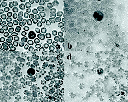
Most of the white cells were either neutrophils or lymphocytes, the
latter being the most abundant. Neutrophils (Figure 1a) were 30±4.5%
of total leukocytes, showed lobulated nuclei (either segmented or
unsegmented) and neutrophilic cytoplasmic granules. The diameter
varied between 10 and 12 μm. Lymphocytes (Figure 1c) were 66±10% and
varied in diameter between 9 and 12 μm, being mostly below 10 μm. The
dense, dark nucleus occupied most of the cell, while the very narrow
band of cytoplasm was light blue. Monocytes (Figure 1b) represented
3±1% and were the largest cells observed with a diameter from 10 to 16
μm. Their distinguishing nucleus was large, indented, resembling a
horse-shoe. The cytoplasm was pale-blue stained and vacuolated.
Platelets were small with an intensively purple-stained central zone.
Eosinophils (Figure 1d) were rare (1%), ranging in diameter from 10 to
14 μm, with a ringed nucleus filling the periphery of the cell. The
cytoplasm was filled with bright pink granules. Basophils were not
observed in the peripheral blood of P. sungorus.
|
|
Figure 1. Blood smears of Djungarian hamster: a) neutrophil b) monocyte c) lymphocyte d) eosinophil (May-Grünwald and Giemsa stain), ×1000 magnification
Click image to enlarge |
The general features of Djungarian hamster blood cells were not
dissimilar from those of the other rodent species. The mean diameter
of the erythroctes (6.02 μm) was smaller than that of
Mesocricetus auratus (6.8 μm) (Chicewicz and Dulemba, 1968)
and Cricetulus griseus (6.8 μm) (Moore, 1966). This seems to
confirm that, among mammals in the same family, smaller species have
smaller erythrocytes (Dunaway and Lewis, 1965).
Howell-Jolly bodies were not detected because in this species the
spleen selectively removes these erythrocytic inclusions (Udroiu,
2007). The relatively high percentage of polychromatic erythrocytes is
similar to that detected in M. auratus (Smith
et al., 2010): this is a common feature among rodents and is
probably due to the relatively short erythrocyte lifespan.
Leukocyte morphology and dimensions were the same as in
M. auratus, the golden hamster. The percentage of neutrophils
and monocytes was slightly higher than in the golden hamster, while
that of lymphocytes was somewhat lower.
The results of this study add new information to our knowledge of
Djungarian hamster haematology and may provide baseline values useful
for veterinarians and biologists dealing with this species both in the
laboratory and in the field.
Acknowledgements
No external funding was received for this study.
References
-
Chicewicz M & J Dulemba: Morfologia krwi chomika
złocistego (Mesocricetus auratus Waterh.). Medycyna
Weterynaryjna, 1968, 12, 746-749.
-
Dunaway PB & LL Lewis: Taxonomic relation of
erythrocyte count, mean corpuscular volume, and bodyweight in
mammals. Nature, 1965, 205, 481-484.
-
Ebling FJ: Photoperiodic differences during development in
the dwarf hamsters Phodopus sungorus and
Phodopus campbelli. Gen Comp Endocrinol, 1994, 95,
475-482.
-
Ebling FJ & P Barrett: The regulation of seasonal
changes in food intake and body weight. J Neuroendocrin, 2008,
20, 827-833.
-
Hoffmann K: Effects of short photoperiods on puberty,
growth and moult in the Djungarian hamster (Phodopus sungorus). J Reprod Fert, 1978, 54, 29-35.
-
Larkin JE, T Yokogawa, HC Heller, P Franken & NF Ruby:
Homeostatic regulation of sleep in arrhythmic Siberian hamsters. Am
J Physiol, 2004, 287, R104-R111.
-
Moore W: Hemogram of the Chinese hamster. Am J Vet Res,
1966, 27, 608-610.
-
Paul MJ, NT George, I Zucker & MP Butler: Photoperiodic
and hormonal influences on fur density and regrowth in 2 hamster
species. Am J Physiol, 2007, 293: R2363-R2369.
-
Ruby NF, T Kang & HC Heller: Melatonin attenuates
photic disruption of circadian rhythms in Siberian hamsters. Am J
Physiol, 1997, 273, R1540-R1549.
-
Ruby NF: Paraventricular nucleus ablation disrupts daily
torpor in Siberian hamsters. Brain Res Bull, 1995, 37,
193-198.
-
Smith SA, KL Zimmerman & DM Moore. In Weis DJ &
Wardrop KJ eds.
Schalm’s veterinary hematology, 6th edition. p
904-909. Blackwell Publishing, Iowa, 2010.
- Udroiu I: A micronucleus test for the Djungarian hamster, Phodopus sungorus, in environmental monitoring. Povolzhskiy J Ecol, 2007, 1, 75-77.