
Original scientific article
Determination of iohexol in canine plasma – strong correlation
between enzyme-linked immunosorbent assay, highperformance
liquid chromatography, and neutron activation
analysis
by VV. Ortín-Piquerasb, e*, T. Spillmannb, M. Pöytäkangasc, D.E. Vaccarod, S. Sankarib, R. Fríasa, e
aCentral Animal Laboratory, University of Turku, Turku, 20520
Finland
bDepartment of Equine and Small Animal Medicine, Faculty of
Veterinary Medicine, University of Helsinki, Helsinki, 00014
Finland
cDepartment of Production Animal Medicine, Faculty of Veterinary
Medicine, University of Helsinki, Helsinki, 00014 Finland
dBioPhysics Assay Laboratory, Inc., Worcester, Massachusetts, 01603
USA
eComparative Medicine, Karolinska Institutet, Stockholm, 171 77
Sweden.
 Correspondence: V. Ortín-Piqueras
Department of Equine and Small Animal Medicine,
Faculty of Veterinary Medicine, University of Helsinki,
Helsinki, 00014 Finland.
Email: victoria.ortinpiqueras@helsinki.fi
Summary
Iohexol is a non-radioactive, iodinated, water-soluble radiographic contrast medium that is widely used in detection imaging for both clinical and scientific purposes. It has also been used as a marker for glomerular filtration rate (GFR) and intestinal permeability (IP) in both humans and animals, such as dogs, rats and cats. Currently, iohexol is determined mainly by high-performance liquid chromatography (HPLC) methods which limit its use in veterinary clinical practice. The aim of this study was to validate an enzyme-linked immunosorbent assay (ELISA) and its accuracy for the measurement of iohexol in canine plasma by comparison with HPLC and neutron activation analysis (NAA). Blank and iohexol-containing blood samples (n=100) from Beagle dogs were collected from the jugular vein in lithium heparin tubes before and after intravenous application of 3.0 g iohexol/dog via the cephalic vein.
The results of this study show that the correlation coefficients when comparing ELISA vs. HPLC (r=0.99), ELISA vs. NAA (r=0.99) and HPLC vs. NAA (r=0.98) are all excellent. In conclusion, the measurement of iohexol from canine plasma using ELISA is as reproducible and reliable as using HPLC or NAA. However, using ELISA for measuring iohexol may be more practical, economical and useful for clinical practice and research than using HPLC or NAA.
Introduction
Iohexol (5-[N-(2,3-dihydroxypropyl) acetamido]-2,4,6-triiodo-N, N’-bis(2,3-dihydroxypropyl) isophthalamide) is a non-radioactive, low-osmolar, iodinated, water-soluble radiographic contrast medium (Andersen et al. 2001). This molecule has been utilized as a practical and reliable marker of glomerular filtration rate (GFR) in humans, pigs, horses, donkeys, dogs, rats and cats (Gleadhill and Michell 1996; Schwartz and Furth 2007; Bexfield et al. 2008; Wang et al. 2012; Meucci et al. 2013; Hellqvist et al. 2015; Meucci et al. 2015; Passos et al. 2015; Schwertner and Weld, 2015). Iohexol has also played a fundamental role in assessing intestinal permeability (IP) (Andersen et al. 2001), having been successfully used as an IP marker in humans, dogs, horses and rats (Halme et al. 1997; Halme et al. 2000; Frías et al. 2009a; Klenner et al. 2009; Koskinen, Hewetson and Pöytakangas 2015).
Although the current gold standard contrast agent for assessment of
GFR and IP is 51Cr-EDTA (Frías, Sankari and Westermarck 2004; Frías et
al. 2009b), there is evidence that iohexol shares a similar IP and GFR
pathway with this marker in dogs and humans, respectively (Klenner et
al. 2009; Slack et al. 2014). However, due to its radioactivity,
51Cr-EDTA is used only by specialized institutions and not in routine
clinical practice (Klenner et al. 2007).
The main detection techniques used for iohexol determination have been
high performance liquid chromatography (HPLC) and neutron activation
analysis (NAA), because the characteristics of these detection methods
for assessment of the iohexol concentration are the best known and
best validated to date (Albert et al. 2003; Soman, Zahir and Akhlaghi
2005; Klenner et al. 2007; Klenner et al. 2009; Pöytäkangas et al.
2010; Gerova et al. 2011; Meucci et al. 2013; Luis-Lima et al. 2014)
A further validation of a practical, reliable, accurate and less
costly method for the determination of iohexol concentrations has been
considered necessary, particularly for the clinical use of this marker
(Frías, Sankari and Westermarck 2004; Klenner et al. 2009; Meucci et
al. 2013; Frías et al. 2014; Jovanović et al. 2015;). Enzyme-linked
immunosorbent assay (ELISA) has been widely used in veterinary
clinical practice and research showing that it is a simple method to
perform and deliver reliable results. However, there are to our
knowledge no publications assessing the validity of an ELISA for
measuring the concentration of iohexol in canine plasma. We tested the
hypothesis that the results of an ELISA for determining iohexol in
canine plasma are well correlated with the results of previously
validated HPLC and NAA methods.
Materials & Methods
Plasma samples
The experimental protocols using dogs were approved by the local
Ethics Committee for Animal Experiments of the University of Helsinki,
Finland (license numbers STU 758 A and STU 776 A). All the dogs were
cared for and used in experiments in accordance with the principles
outlined in the Finnish and European legislation on the use of
vertebrate animals for scientific purposes (European Community Council
Directive 86/609/EEC, Council of Europe, 1986; Finnish Government,
1985; Finnish Government, 1996).
The plasma samples (n = 100) taken from 10 clinically healthy
Beagle dogs were historical samples from an unrelated study in which
iohexol had been used to determine GFR (Pöytäkangas et al. 2010). The
dogs were maintained in indoor pens, spending about 4 h daily in
outdoor runs. The environmental temperature indoors was maintained
within a range of c.15-24oC. Prior to commencement of the study, each
dog was subjected to a clinical examination and a complete plasma
biochemical analysis and blood count to confirm the absence of
disease.
The blood samples were collected from the jugular vein in lithium
heparin tubes before and after intravenous application of 3.0 g
iohexol/dog (Omnipaque 300 mg mL-1; GE Healthcare, Helsinki, Finland)
via the cephalic vein (Pöytäkangas et al. 2010). Subsequently, blood
samples were kept on ice packs until being centrifuged (1,300 x g for
10 min). Plasma was drawn off and frozen at -20oC. Prior to analysis,
plasma samples were thawed to room temperature and mixed thoroughly.
All samples were analyzed in duplicate (Pöytäkangas et al. 2010).
Iohexol concentration was analyzed by rapid high-performance liquid
chromatography-ultraviolet (LC-UV) in the Faculty of Veterinary
Medicine, Helsinki, Finland, prior shipping the samples to BioPhysics
Assay Laboratory Inc (BioPAL, Worcester, MA, USA) for analysis using
ELISA and NAA.
Immunoassay analysis
The ELISA (functional immunoassay technology (FIT)-GFRTM Iohexol kit,
BioPAL, Worcester, MA, USA) was used according to the
manufacturer’s instructions. Iohexol standards were prepared
using the ELISA kit diluent (0.01, 0.03, 0.1, 0.3, 1.0, 3.0, 10.0
μg/mL), and then, to bring the samples within the active range of the
standard curve, the plasma samples were diluted 1:300.
Fifty μL of standard or diluted sample were pipetted into wells of a
96-well coated plate, and then 50 μL of rabbit anti-iohexol were added
to each well. The plate was incubated on an orbital shaker for 1 h,
and immediately washed 3 times with a Tween 20 PBS wash buffer (Elx50
WasherBiotek Instruments, Inc., Winooski, VT, USA). Then, 100 μL of
goat anti-rabbit IgG-HRP was pipetted into each well and again
incubated for 30 min followed by a second plate wash cycle. Substrate
reagent (100 μL) was added to each well and incubated for 30 min at
room temperature. Finally, stop reagent (100 μL) was added to each
well.
The absorbance at 450 nm was recorded using software supplied with the
plate reader (Multiskan® Spectrum, Thermo Electron Corporation,
Waltham, MA, USA), and data from the standards were fitted to a
4-parameter logistic function. By interpolation, the concentration of
iohexol present in each sample was determined.
Neutron Activation Analysis
Each plasma sample was centrifuged and 100 μL of plasma was
transferred to a vial designed for neutron activation analysis (NAA),
as described previously (Albert et al. 2003; Mandelbrot et al. 2007).
Each vial contained a known amount of a metallic monitor to account
for potential neutron-flux density variations during neutron
activation (Reinhardt et al. 2001). All vials were activated by
exposure to a field of neutrons. These vials were stored for 48 h to
allow the short-lived activation products to decay. The concentration
of the resultant radioactive nuclei in each vial was then measured by
spectrographic analysis (Reinhardt et al. 2001). Iohexol standards
were prepared in the same way.
Rapid high-performance liquid chromatography-ultraviolet
Iohexol concentration in canine plasma samples was determined using
rapid HPLC-UV previously developed and validated for the assessment of
the GFR and IP (Pöytäkangas et al. 2010). In brief, trifluoracetic
acid (TFA) was used for protein precipitation and iohexol extraction
from canine plasma, followed by vortex mixing and centrifugation.
4-Aminobenzoic acid (para-aminobenzoic acid, PABA) was added as an
internal standard. Samples were analyzed by an Agilent model 1200
series rapid resolution LC system (Agilent Technologies, Waldbronn,
Germany). The mobile phase gradient was linear and consisted of
methanol and water (pH 3.0, adjusted with TFA). Gradient stop time was
8 min and post-time 5 min. The flow rate was 1 mL/min. At a column
oven temperature of 50oC the LC operating pressure was approximately
310 bar. Iohexol detection was carried out at a wavelength of 246 nm,
and then using a Chemstation data system the results were calculated
(Pöytäkangas et al. 2010).
Statistical methods
For each sample, the iohexol concentration measured by ELISA was
compared to the concentration measured by NAA and HPLC using Bland and
Altman analysis (Bland and Altman 1986). NAA, ELISA and HPLC were
compared by calculating correlation coefficient, precision (standard
deviation of bias), bias (the difference between a population mean of
the measurements), and accuracy (absence of bias, percent error from
the true value) (Albert et al. 2003; Walther and Moore 2005).
Statistical analysis was performed by using GraphPad Prism 6.07
(GraphPad Software Inc., La Jolla, CA, USA).
|
|
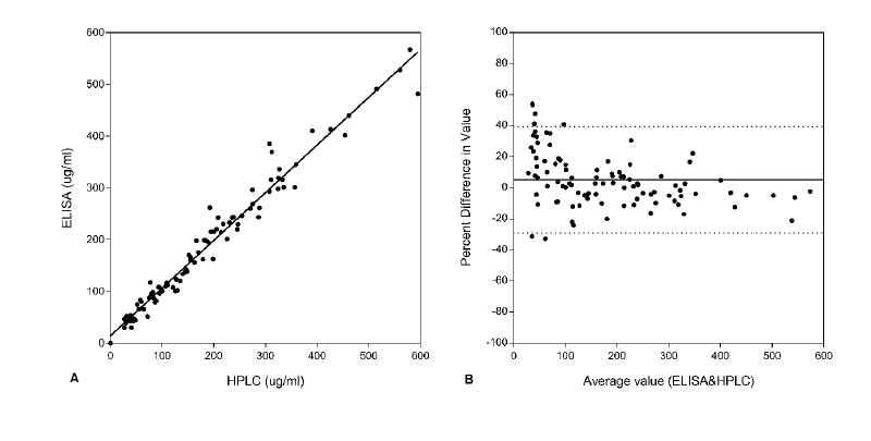
Figure 1. The iohexol concentration (μg/mL) in
collected plasma samples (n = 100) measured by ELISA and NAA. A.
ELISA values compared directly with NAA values. y = 1.002x +
7.164; r = 0.99 (p <0.0001). B. Difference against the mean
iohexol value. Click image to enlarge |

Results
No evidence of gastrointestinal or renal disease was found in any of the dogs used in the study. All dogs were in good body condition, no abnormalities were identified on clinical examination, plasma biochemical analysis and blood count. Iohexol tolerance was excellent in the dogs and there were no adverse effects.
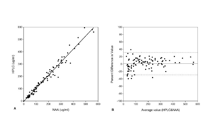
The range of iohexol concentrations measured with the ELISA was 0–600 µg/mL. The correlation coefficients when comparing ELISA vs. HPLC (r=0.99), ELISA vs. NAA (r=0.99), and HPLC vs. NAA (r=0.98) were all strong (Figs 1A, 2A & 3A). For the direct comparison between ELISA and NAA, the bias was 6.52 μg/mL, the precision was 10.42 μg/mL, and the accuracy was such that 97% of the measured values were within 2 standard deviations (SD) of the mean (Fig. 1B). For the comparison between ELISA and HPLC, the bias was -5.123 μg/mL, the precision was 17.5 μg/mL, and the accuracy was such that 94% of the measured values were within 2 SD of the mean (Fig. 2B). Finally, in the comparison between HPLC and NAA, the bias was -1.36 μg/mL, the precision was 15.55 μg/mL, and the accuracy was such that 94% of the measured values were within 2 SD of the mean (Fig. 3B).
|
|
Figure 2. The iohexol concentration (μg/mL) in
collected plasma samples (n = 100) measured by ELISA and HPLC.
A. ELISA values are compared directly with HPLC values y =
0.9218x + 14.04; r = 0.99 (p <0.0001). B. Difference against
the mean iohexol value. Click image to enlarge |
|
|
Figure 3. The iohexol concentration (μg/mL) in
collected plasma samples (n = 100) measured by HPLC and NAA. A.
HPLC values are compared directly with NAA values y = 1.062x –
3.273; r = 0.98 (p <0.0001) B. Difference against the mean
iohexol value. Click image to enlarge |
Discussion
In view of the excellent correlation between ELISA, HPLC and NAA, these methods may be considered comparable detection techniques for the determination of iohexol concentration in canine plasma. However, HPLC and NAA have many disadvantages. Although HPLC is a fast, reliable, and the most widely used technique for analysis of iohexol (McMaster 2007; Pöytäkangas et al. 2010; Meucci et al. 2013) its cost and technical complexity may make it impractical to use for clinical research and as a routine detection test for assessing renal function in patients (Rocco et al. 1996; McMaster 2007; Slack et al. 2014). NAA has advantages as a reproducible technique for detecting iohexol (Albert et al. 2003; Mandelbrot et al. 2007). However, the requirement of a nuclear reactor to work with radioactive materials and specific licenses make it an unsuitable method for routine clinical diagnostics or research projects (Albert et al. 2003; Loveland, Morrissey and Seaborg 2005; De Groot 2008). The main benefits of the ELISA method are its high sensitivity, high sample throughput, short time of analysis and relatively low cost when compared to HPLC and NAA (Hendrikje et al. 1997; Khan et al. 1998).
ELISA would have a greater application in clinical and experimental
research not only for being a non-radioactive method, but also for
being quick and easy to perform even for non-specialists, less
technically demanding and allowing the use of a small sample size
(Hendrikje et al. 1997; Khan et al. 1998; Alamdari et al. 2005).
Moreover, because iohexol is a clinically accessible reagent, and as
the ELISA reagents are widely available and provided in kit format,
this approach may enable identical results and better standardization
to be achieved among core laboratories and independent investigators.
The validation of new diagnostic techniques for analysis of iohexol
concentration in canine plasma facilitates the assessment of GFR and
IP in dogs, revealing that ELISA is as reproducible and reliable as
HPLC or NAA. When comparing the advantages and disadvantages of the
different methods, ELISA may be more economical and accessible for
clinical practice and research than HPLC or NAA. In addition, a simple
and reliable method for the determination of iohexol as a
non-radioactive marker of GFR and IP may help not only veterinary
clinicians to diagnose renal and intestinal dysfunction but also
researchers to better investigate the role of the kidney and
intestinal integrity in different canine disease models.
Acknowledgements
We are grateful to Juhana M. Honkavaara and Marja Raekallio for providing the plasma samples.
References
- Alamdari, D.H., Kostidou, E., Paletas, K., Sarigianni, M., Konstas, A.G., Karapiperidou, A., Koliakos, G., (2005). High sensitivity enzyme-linked immunosorbent assay (ELISA) method for measuring protein carbonyl in samples with low amounts of protein. Free Radical Biology and Medicine. 39, 1362-1367.
- Albert, D.A., Cohen, A.J., Mandelbrot, D.A., Reinhardt, C.P., Dickson, E.W., (2003). Neutron-activation analysis: A novel method for the assay of iohexol. Journal of Laboratory and Clinical Medicine. 141, 106-109.
- Andersen, R., Stordahl, A., Aase, S., Laerum, F., (2001). Intestinal permeability of x-ray contrast media iodixanol and iohexol during bacterial overgrowth of small intestines in rats. Digestive Diseases and Sciences. 46, 208–213.
- Bexfield, N.H., Heiene, R., Gerritsen, R.J., Risøen, U., Eliassen, K.A., Herrtage, M.E., Michell, A.R., (2008). Glomerular filtration rate estimated by 3-sample plasma clearance of iohexol in 118 healthy dogs. Journal of Veterinary Internal Medicine. 22, 66-73.
- Bland, J.M., Altman, D.G., (1986). Statistical methods for assessing agreement between two methods of clinical measurements. Lancet. 1, 307-310.
- De Groot, P.A., (2008). Handbook of Stable Isotope Analytical Techniques. In: De Groot, P.A. (Ed.). 1st Edn. Elsevier Science, Vol.2.
- Frías, R., Sankari, S., Westermarck, E., (2004). 51Cr-EDTA absorption blood test: an easy method for assessing small intestinal permeability in dogs. Journal of Veterinary Internal Medicine. 18, 156-159.
- Frías, R., Ouwehand, A., Spillmann, T., Salminen, S., Gueimonde, M., (2009a). Comparison of 51Cr-EDTA and iohexol as blood markers for intestinal permeability testing in Beagle dogs. The Veterinary Journal. 192, 123-125.
- Frías, R., Ouwehand, A.C., Spillman, T., Vankerckhovend, V., Hewicker-Trautweine, M., Salminenc, S., Gueimonde, M., (2009b). Effect of clinical and probiotic Lactobacillus rhamnosus strains on intestinal permeability and bacterial translocation in healthy and colitic rats. Food Research International. 42, 636-640.
- Frías, R., Ouwehand, A.C., Jaakkola, U-M., Spillman, T., Gueimonde, M., (2014). An in vivo permeability test protocol using iohexol to reduce and refine the use of laboratory rats in intestinal damage assessment. Scandinavian Journal of Laboratory Animal Science. 40, 1-6.
- Gerova, V.A., Stoynov, S.G., Katsarov, D.S., Svinarov, D.A., (2011). Increased intestinal permeability in inflammatory bowel diseases assessed by iohexol test. World Journal of Gastroenterology. 17, 2211-2215.
- Gleadhill, A., Michell, A.R., (1996). Evaluation of iohexol as a marker for the clinical measurement of glomerular filtration rate in dogs. Research in Veterinary Science. 60, 117-121.
- Halme, L., Edgren, J., Turpeinen, U., Von Smitten, K., Stenman, U.H., (1997). Urinary excretion of iohexol as a marker of disease activity in patients with inflammatory bowel disease. Scandinavian Journal of Gastroenterology. 32, 148-152.
- Halme, L., Turunen, U., Tuominen, J., Forsstrom, T., Turpeinen, U., (2000). Comparison of iohexol and lactulose-mannitol tests as markers of disease activity in patients with inflammatory bowel disease. Scandinavian Journal of Clinical and Laboratory Investigation.60, 695-701.
- Hellqvist, A., Heine, R., De Baere, S, Croubels, S, Hedeland, Y., (2015). Development of a capillary electrophoretic method for determination of plasma clearance of iohexol in dogs and cats. Biomedical Chromatography. 29, 504-513.
- Hendrikje, B., Chan, T.P., Sluis, K.B., Domigan, N.M., Winterbourn, C.C., (1997). Protein Carbonyl Measurement by a Sensitive ELISA Method. Free Radical Biology & Medicine 23, 361-6.
- Jovanović, M., Rakić, T., Tumpa, A., Jančić Stojanović, B., (2015). Quality by Design approach in the development of hydrophilic interaction liquid chromatographic method for the analysis of iohexol and its impurities. Journal of Pharmaceutical and Biomedical Analysis. 110, 42-48.
- Khan, J., Brennand, D.M., Bradley, N., Gao, B., Bruckdorfer, R., Jacobs, M., (1998). 3-Nitrotyrosine in the proteins of human plasma determined by an ELISA method. Biochemical Journal. 330, 795-801.
- Klenner, S., Bergmann, C., Strube, K., Ternes, W., Spillmann, T., (2007). SPE for endo- and exo-iohexol analysis with HPLC in canine serum and rat urine. Chromatographia. 65, 733-736.
- Klenner, S., Frías, R., Coenen, M., Failing, K., Hewicker-Trautwein, M., Ternes, W., Verspohl, J., Spillmann, T., (2009). Estimation of intestinal permeability in healthy dogs using the contrast medium iohexol. Veterinary Clinical Pathology. 38, 353-360.
- Koskinen, M. J., Hewetson, M., Pöytakangas, M.R., (2015). Iohexol as a marker of intestinal permeability in the horse. Equine Veterinary Journal. 48, 1-8.
- Loveland, W.D., Morrissey, D.J., Seaborg, G.T., (2005). Modern Nuclear Chemistry. 1st Edn. Wiley-Interscience, N.Y., pp. 704.
- Luis-Lima, S., Gaspari, F., Porrini, E., García-González, M., Batista, N., Bosa-Ojeda, F., Oramas, J., Carrara, F., González-Posada, J.M., Marrero, D., Salido, E., Torres, A., Jiménez-Sosa, A., (2014). Measurement of glomerular filtration rate: Internal and external validations of the iohexol plasma clearance technique by HPLC. Clinica Chimica Acta. 430, 84-85.
- Mandelbrot, D.A., Dhaliwal, S.K., Evan, N.R., Licho, R., Reinhardt, C.P., Jaffry, S., (2007). Validation of neutron activation as a novel method to determine glomerular filtration rate. Nephron Clinical Practice. 107, 117-122.
- McMaster, M.C., (2007). HPLC: a practical user’s guide. 2nd edn. Wiley-Interscience, N.Y. A John Wiley & Sons, Inc., Publication.
- Meucci, V., Guidi, G., Melanie, P., Breghi, G., Lippi, I., (2013). A limited sampling, simple, and useful method for determination of glomerular filtration rate in cats by using a new accurate HPLC method to measure iohexol plasmatic concentrations. Journal of Veterinary Medicine [Online]. Available from: http://dx.doi.org/10.1155/2013/569121
- Meucci, V., Sgorbini, M., Bonelli, F., Corazza, M., Lippi, I., Intorre, L., Guidi, G., (2015). Determination of Glomerular Filtration Rate in Adult Horses and Donkeys by Single IV Administration of Iohexol. Journal of Equine Veterinary Science. 351, 36-40.
- Passos, M.T., Nishida, S.K., Câmara, N.O.S., Shimizu, M.H., Mastroianni-Kirsztajn, G., (2015). Iohexol Clearance for Determination of Glomerular Filtration Rate in Rats Induced to Acute Renal Failure. PLOS One [Online]. Available from: https://doi.org/10.1371/journal.pone.0123753
- Pöytäkangas, M., Saario-Paunio, E., Putkonen, T., Saastamoinen, I., Frías, R., Spillmann, T., Saloniemi, H., (2010). Rapid LC-UV analysis of iohexol in canine plasma for glomerular filtration rate determination. Chromatographia. 71, 211-216.
- Reinhardt, C.P., Dalhberg, S., Tries M.A., Marcel, R., Leppo, J.A., (2001). Stable labeled microspheres to measure perfusion: validation of a neutron activation assay technique. American Journal of Physiology-Heart and Circulatory Physiology. 280, 108-116.
-
Rocco, M.V., Buckalew V.M., Moore, L.C., Shihabi, Z.K., (1996).
Capillary electrophoresis for the determination of glomerular
filtration rate using nonradioactive iohexol.
American Journal of Kidney Diseases. 28,
173-177.
Schwartz, G.J., Furth, S.L., (2007). Glomerular filtration rate measurement and estimation in chronic kidney disease. Pediatric Nephrology. 22, 1839–48. - Schwertner, H.A., Weld, K.J., (2015). High-Performance Liquid- Chromatographic Analysis of plasma iohexol concentrations. Journal of Chromatographic Science. 53, 1475-1480.
- Slack, A., Tredger, M., Brown, N., Corcoran, B., Moore, K., (2014). Application of an isocratic methanol-based HPLC method for the determination of iohexol concentrations and glomerular filtration rate in patients with cirrhosis. Annals of Clinical Biochemistry.51, 80-88.
- Soman, R.S., Zahir, H., Akhlaghi, F., (2005). Development and validation of an HPLC-UV method for determination of iohexol in human plasma. Journal of Chromatography B. 816, 339-343.
- Wang, E., Meier, D.J., Sandoval, R.M., von Hendy-Willson, V.E., Pressler, B.M., Bunch, R.M., Alloosh, M., Sturek, M.S., Schwartz, G.J., Molitoris, B.A., (2012). A portable fiberoptic ratiometric fluorescence analyzer provides rapid point-of-care determination of glomerular filtration rate in large animals. Kidney international. 81, 112-117.
- Walther, B.A., Moore, J.L., (2005). The concepts of bias, precision and accuracy, and their use in testing the performance of species richness estimators, with a literature review of estimator performance. Ecography. 28, 815-829.