
Introduction
Urethral strictures remain a serious challenge in mod-ern
urology (King and Rouke 2019). Traditional treat-ments such as
urethral dilatation, primary anastomosis, substitution
urethroplasties often fall short of achiev-ing long-term success
(Güller 2021), with outcomes that may not always meet the
desired expectations (Lu-men 2009). Bioengineering holds promise
for discov-ering new treatment methods, with artificial
biological tissues offering a potential breakthrough in
restoring damaged urethras (Chapple 2020).
Currently, a lot of laboratory research is under-way to create
the right biologically relevant tissue that can be used in
clinical practice. However, these stud-ies require testing in
animals before experiments can progress to trials in humans. To
solve the issues of formation and treatment of urethral
strictures, first, a stricture of the urethra is induced in
animals, and then methods of treating are studied (Horiguchi et
al. 2021).
An appropriate animal model of urethral stric-ture is needed for
further in-depth investigation of the mechanisms of stricture
formation and treatment. Rab-bit and dog models of urethral
stricture have recently been produced by open surgery,
electroresection, elec-trocoagulation (Meria et. al. 1999),
laser ablation (Hu et al. 2014) and pharmaceutical agent
induction methods (Hua et al. 2018). Animal models created by
open sur-gery have desirable reproducibility because of precise
control of the procedure, but open surgery may result in severe
wounds and has a higher incidence of urethral fistula formation
(Yao et al. 2022).
Most of the studies on the development of ure-thral stricture
patterns are limited to the final results, indicating whether
the urethral stricture was formed successfully, what was its
length and depth, how many animals survived (Xue et al. 2016).
Unfortunately, there is lack of data on how the animals felt
during the exper-iment and the challenges that researchers
faced. These features are very important, especially considering
that the animals with urethral strictures will have to
partic-ipate in further studies when the strictures are treated
with artificial tissues. Pain, stress, and fatigue are com-mon
to many animals, including rabbits. Controlling pain and stress
improves animal well-being and postop-erative outcomes (Wenger
2012). In rabbits, pain recog-nition can be particularly
challenging because, as with many other prey species, these
animals are predisposed to mask any sign of pain (Benato et al.
2019).
There is evidence that pain causes a decrease in activity and
nutrition in rabbits (DiVincenti et al. 2016). Since rabbits’
metabolism is geared to a constant sup-ply of nutrients from the
digestive tract, a decreased or absent food intake and the
subsequent mobilization of fat reserves can lead to ketoacidosis
and hepatic li-pidosis (Varga 2013). Weight loss is known to
contrib-ute to complications such as increased susceptibility to
infections, delayed wound healing, and in severe cases,
mortality (DiVincenti et al. 2016).It is important to pay
attention to the behavio-ral changes of rabbits postoperatively
(Gargiulo et al. 2025). One of the main behavioral features
presenting when rabbits respond to pain is freezing and distress
by remaining motionless, especially in the presence of an
observer (Johnston 2005).
The purpose of our pilot study was to evaluate the welfare of
rabbits after the induction of urethral stric-ture, with the
intention of preparing the animals for further experiments.
Rabbits must be in good health before they can participate in
further studies on the treatment of urethral strictures. This
was the initial phase of a larger project, aiming to compare
different urethral stricture treatment methods.
Materials and Methods
Four New Zealand white rabbits (Oryctolagus cuniculus)
(Animalab, Hungary Kft.) were selected in this pilot study. The
average age of rabbits was 12 months, the average weight was
4465 g.
They were randomly divided into two study groups, each
consisting of 2 animals. Group A was assigned for a simple open
urethrotomy, group B was assigned for open electrocauterization
of the urethral endothelium to induce stricture formation.
During surgery, rabbits were anesthetized by in-tramuscular
injection of ketamine hydrochloride (35 mg/kg, bioketane,
vetoquinol) and xylazine (5 mg/kg, Xylamedo, Bimeda).
Intraoperative monitoring of animal pulse and oxygenation was
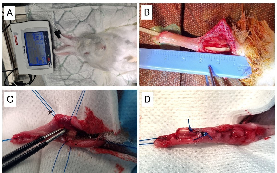
caried out. (Figure 1A) The operative field in the perineal area
was steri-lized with Povidone-iodine solution. A single
intramus-cular injection of the antibiotic Enrofloxacin 2.5
mg/kg (Bayer B.V.) was performed to prevent infections, and the
non-steroidal analgesic Carprofen 4.0 mg/kg (Rycarfa, KRKA) was
administered intramuscularly to relieve postoperative pain.
In both groups, a ventral incision of the middle part of the
penile urethra was performed under general anesthesia (Figure
1B). In group A, the urethral lumen was opened for a length of 2
cm. A 10 Ch Nelaton type catheter was inserted. The wound was
closed with two layers of Vicryl rapid 4-0 sutures, suturing
subcu-taneous tissue and skin, but leaving the edges of the
urethral endothelium free of sutures. As a result, the urethral
defect was left not repaired, and a stricture is expected to
form. The catheter was sutured to the fore-skin with two 3-0 PDS
sutures to remain in place for 5 days (Figure 1D).
In rabbits of group B the same ventral incision of the middle
part urethra was made. After opening the lumen, the endothelium
of the urethra was super-ficially electrocauterized using
bipolar electrocautery, avoiding perforation and damage to the
surrounding tissues. (Figure 1C) The wound was closed in the
same manner as in group A, leaving a 10 Ch Nelaton catheter
indwelling
The rabbits were housed individually in a
tempera-ture-controlled cage (TechnoPlast) with a humidity of
50-55 % for a 12-hour light-dark cycle and had freeaccess to
chow (Maintenance Diet for Rabbits, Altro-min) and tap water.
During the first week after surgery,the rabbits’ wounds were
cared daily and, later, weekly.
Urethral catheters were secured to the foreskin and shortened to
the level of the foreskin to minimize the likelihood of
dislodgement. Nevertheless, most catheters were lost or actively
removed by the rabbits within the first two postoperative days.
Even though rabbits have lost catheters they did not experience
uri-nary retentions. There was mild hematuria seen during first
3 days postoperatively.
The rabbits were cared for by a qualified veterinar-ian. To
standardize the follow-up procedure, we used the scale of
physical and behavioral symptoms. The scale consisted of these
parameters: wound condition, behavioral manner, fur quality,
urination quality, nutri-tion quantity. Each parameter was
evaluated in scale 0-1-2, meaning: 0 – poor, 1 – average, 2 –
good. To-tal sum of parameters showed general level of
rabbits’welfare during postoperative follow-up period.
The rabbits were weighed once per week; the ure-thral wound was
disinfected and evaluated periodically. The body temperature was
measured once per week at the inner surface of the ear using an
appropriate non-contact infrared thermometer.
After 4 and 12 weeks, urethrograms were per-formed under general
anesthesia. It was performed in the same manner as initial
surgery by intramuscular administration of Ketamine
hydrochloride (35 mg/kg) and Xylazine (5 mg/kg). The rabbit was
placed on its back, the perineal area was disinfected with
Octisept (octenidine dihydrochloride/phenoxyethanol, Schülke &
Mayr GmbH) spray. The penis was stretched in the caudal
direction, the 8fr catheter inserted into the ure-thra
aproximatelly 2cm, positioned at the site of the urethroplasty
and fixed with a Vicril 2/0 thread. Uro-grafin® (76%, Sodium
Amidotrizoate / Amidotrizo-ate Meglumine, Bayer B.V.) solution
was diluted with sterile saline to 38% and injected through the
cathe-ter. Radiographs were captured by the digital Madical
ECONET mex+100 X-ray equipment, 50kV, 2.0sec/mAs, 100mA mode.
12 weeks after urethral stricture induction the rabbits were
sacrificed and the urethra was dissected. The rabbit penis was
cut at the root part and fixed in 10% formalin solution. The
formalin-fixed samples were embedded in paraffin, sectioned
every 5 mm and stained with hematoxylin and eosin. Samples were
analyzed under a bright-field microscope using a Leica M205C
stereomicroscope at 0.78 times magnification and an Olympus AX70
microscope with 2x and 10x magnification objectives. All
histological specimens were blinded and evaluated by the same
pathologist
Statistical analysis
Due to the small sample size, appropriate statistical analysis was not performed. Analysis of the study results was limited to descriptive statistics, reporting absolute data values and medians. A larger cohort would have increased statistical power and perhaps provided more reliable comparisons between groups. However, even with the limited sample size, the apparent differences between groups strongly support our main conclusion, therefore it was decided not to increase the sample size until the study protocol could be improved.
Results
The average surgery time was 23.75 minutes. The duration of surgery was longer in electrocauterization (B) group (25.00 minutes) compared to simple incision (A) group (22.50 minutes). All 4 rabbits survived the surgery fluently and no intraoperative complications occurred. The rabbits were observed for 6 hours after surgery until they fully woke up from anesthesia. The rabbits were active and showed no signs of illness. Based on previous experimental studies and a review of the literature, no additional analgesia or antibiotics were administered. Apart from the nutritional problems of group B animals described in the weight change and welfare paragraphs, all animals survived to the last stage of the experiment. Minimal blood was found on the bedding on the first postoperative day, which was considered normal postoperative course. Although the urethral catheter was planned to be kept for 5 days, the rabbits removed the catheters from the urethra already on the first postoperative day.
Urethrograms
Urethrograms were performed after 4 and 12 weeks of follow-up.
Under general anesthesia, Urographin 38% solution was instilled
into the urethra, clearly reveal-ing its lumen architecture.
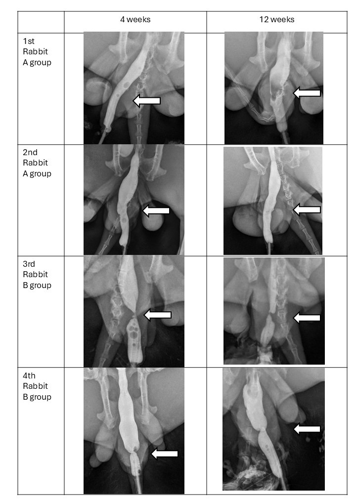
Urethrograms did not show any extravasation or urethral fistulas
in the animals of both groups. The iodine-based contrast
solution easily passed the site of previous surgery and filled
the blad-der. It did not reveal any side effects or reactions
from the rabbit.
In both animals of A group, no signs of urethral stricture were
detected either at 4-week urethrogram or at 12-week urethrogram.
(Figure 2, A. and B.) Other-wise, in both animals of B group the
urethral stricture was clearly detected at 4-week as well as at
12-week urethrograms (Figure 2, C. and D.) Urethral stricture
retained the same size when comparing urethrograms at 4 and 12
weeks.
Histology
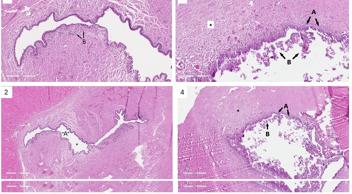
Final histological examination revealed a difference between the
two methods of stricture induction. Although urethral incision
appears to be a serious injury and severe strictures would be
expected in humans, complete recovery was confirmed in rabbits.
The urethra had a normal lumen and was lined by normal
transitional urothelium. Mild infiltration with
polymorphonuclear leucocytes was observed in the urethral wall.
The pathologist did not observe any evidence of fibrosis or
stricture. (Figure 3.1 and Figure 3.2)
Clearly different findings were obtained in samples from
electrocauterization (B) group animals. In this group, serious
damage to the urethra was detected, and a proper stricture was
formed after 12 weeks. The histological findings replicated the
results we had observed during the urethrograms. The surgical
site was fibrotic, the lumen of the urethra was narrow, covered
with squamosal epithelium, which indicates poor wound healing.
Inflammatory infiltration of the urethral wall was observed,
with abundant neutrophils and fibroblasts present within the
tissue. Inflammatory elements were also found in the urethral
lumen. (Figure 3.3 and Figure 3.4)
Histological findings showed notable difference between two
surgical methods – simple incision versus incision and
electrocauterization of the urethral wall. The second method
showed severe lesion to the urethra, generating stable urethral
stricture after 3 months.
Welfare
The experiment aimed to evaluate the welfare of rabbits during
follow-up period. We used the standardized scale of welfare
symptoms, that was described in methodology. The measurements of
temperature, weight, blood tests, and urine samples supported
our data.
One rabbit of incision (A) group had mild postoperative
hematuria, and a swollen wound 1 week after surgery. These
changes disappeared naturally within the second postoperative
week. One rabbit in electrocauterization (B) group had nutrition
difficulties for 3 days postoperatively and needed supportive
nutrition with syringe. Finaly in 4th day the rabbit started
eating and drinking normally. The wound of mentioned group B
rabbit was suspicious on 12th week postoperative, and it was
confirmed as prompt stricture after final autopsy.
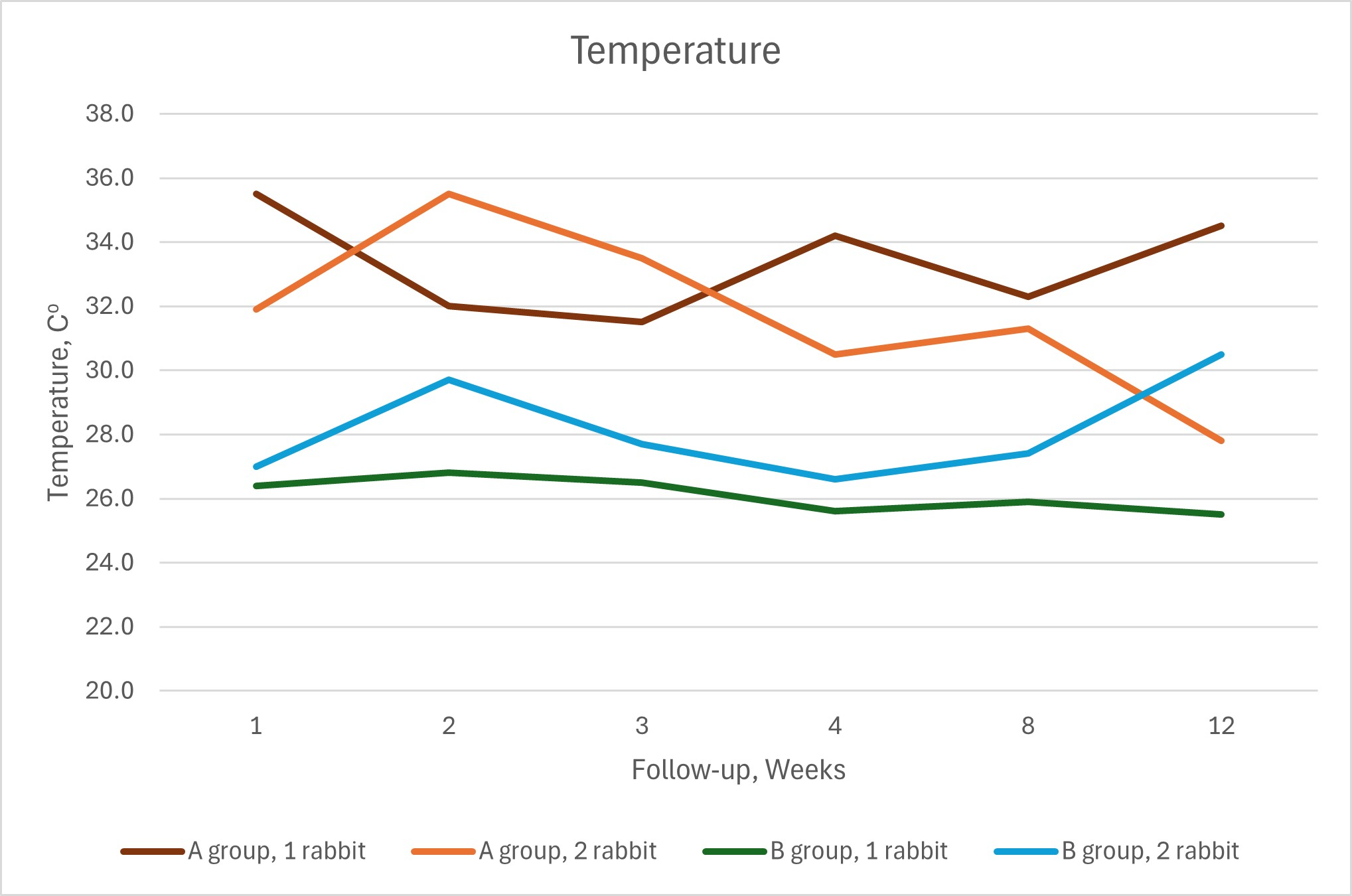
Temperatures of group A rabbits were markedly higher comparing
to the B group rabbits. Otherwise the temperatures of all
rabbits was constantly stable through all experiment time and
did not change indicating any inflammation or other reactions
(Figure 4).
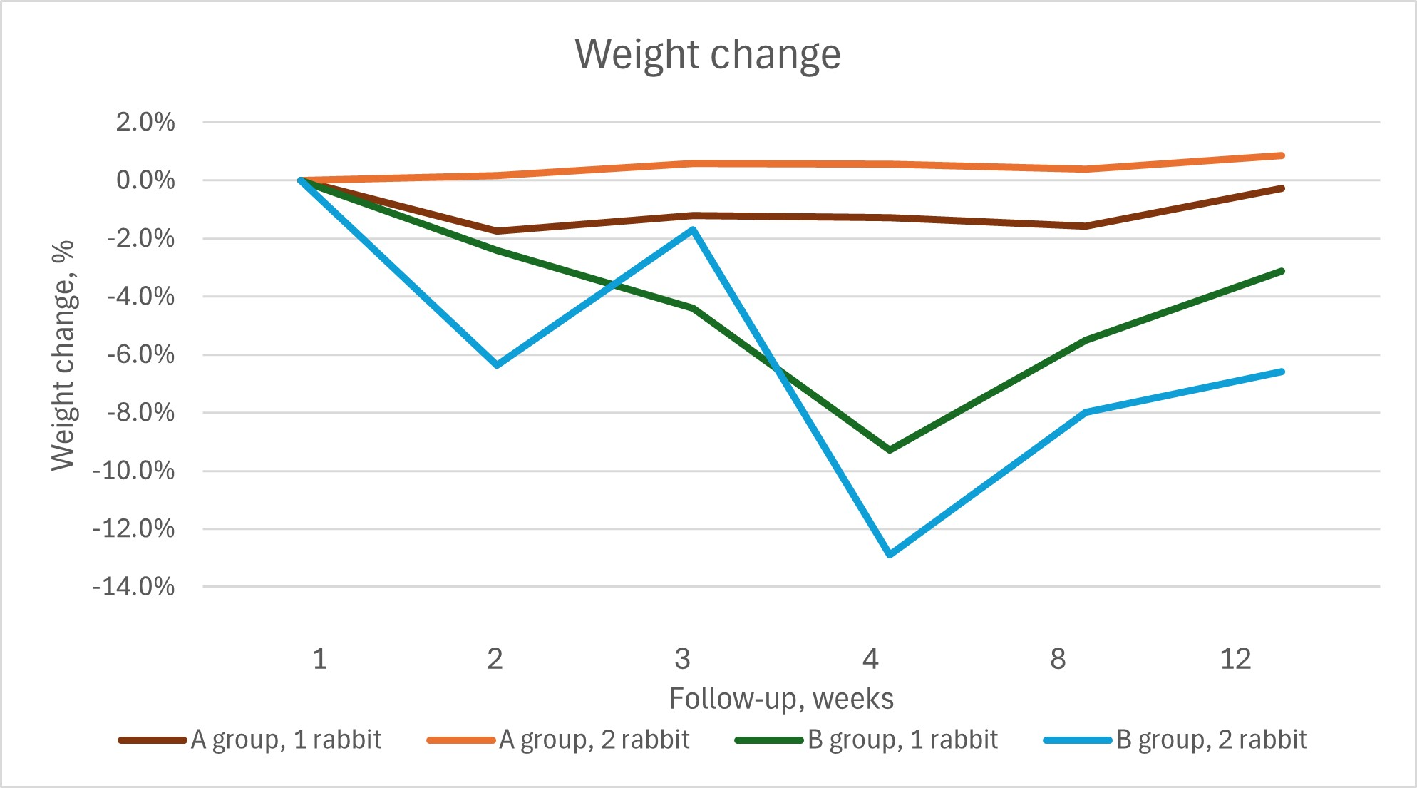
Weight change is one of the most typical indicators of animal
welfare. When experiencing stress, pain or illness, animals
change their eating habits and lose weight (Benato et al. 2019).
This was also seen in the rabbits in this experiment. Rabbits
with more extensive urethral lesions lose a greater percentage
of weight compared to their associates. A reduction in body
weight was observed over the first 4 weeks, followed by a
gradual recovery. This was most relevant for the electrocaut
erization (B) group animals (Figure 5).
Clinical signs like fur change, habits change, urination manner,
wound or hematuria signs did not express any considerable
difference between A and B group animals.
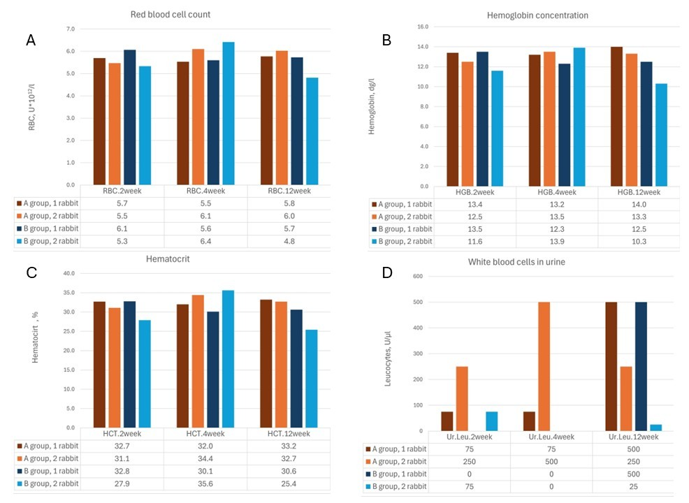
Blood and urine tests were performed at 2, 4 and 12 weeks of
follow-up. Slight decreases in some blood parameters were
observed in electrocauterization (B) group animals at the 12th
week of the experiment. Red blood cell count (RBC), hemoglobin
(HGB) and hematocrit (HCT) levels were reduced in group B
rabbits. In urine sample - leukocyturia varied and did not
differ between A and B groups (Figure 6).
Discussion
The aim of the study was to identify a suitable method for
inducing urethral strictures for future artificial tissue
research, with careful consideration of laboratory animal
welfare. Electrocauterization models tended to develop stable
urethral strictures in previous studies, but the long-term
results needed to be proven (Meria et al. 1999). Effective
animal models for the creation of urethral stricture are needed
to allow urethral reconstruction studies, but appropriate
methods are still under investigation (Faydaci et al. 2012).
This study confirmed that electrocauterization reliably induced
urethral stricture in the electrocauterization (B) group, as
evidenced by urethrograms at both 4 and 12 weeks. By 4 weeks,
the stricture was fully formed and remained stable through week
12. Histological examination supported these findings, revealing
fibrosis of the urethral wall, lumen narrowing, and epithelial
damage, indicating the formation of a true long-term stricture.
In contrast, no stricture was observed in the incision (A) group
at either time point. Our study, which observed the rabbits for
a longer duration compared to others, demonstrated that
electrocauterization in the B group is a reliable method for
inducing urethral stricture. The desired stable outcome was
achieved by 4 weeks and additional time was not necessary for
the development of a tight, firm stricture.
Based on previous studies (Nikolavsky et al. 2016), we aimed to
catheterize the bladder of rabbits postoperatively to prevent
urinary retention or fistula formation. Although 10 Ch Nelaton
catheters were attached with sutures to the foreskin and
shortened to the tip of the penis, the rabbits removed them as
soon as they recovered from anesthesia on the first
postoperative day. It is noteworthy that despite this, there was
no urinary retention or other early postoperative complications.
Some studies suggest that it is not worth leaving a catheter in
the urethra after surgery (Micol et al. 2012). As we observed,
the catheter did not improve the postoperative condition, so we
believe that a catheter is not necessary after open urethrotomy
in rabbits.
The welfare of rabbits after the induction of urethral stricture
is of great importance. This is important not only for the sake
of good laboratory practice (Directive 2010/63/EU; Srinivasan et
al. 2021), but also because these rabbits will be involved in
subsequent experiments related to the treatment of the induced
stricture.
Body weight can be monitored as a percentage change from values
recorded immediately before stricture induction and measuring it
periodically after the operation. According to the data of our
study, despite the strictures formed in the rabbits of
electrocauterization group, serious complications were avoided -
all the rabbits survived, without fistulas, abscesses or urinary
retentions, the rabbits urinated until the end of the
experiment.
However, rabbits in the electrocauterization (B) group consumed
less food and lost more body weight during the first
postoperative weeks compared with the incision (A) group.
Reduced food and water consumption, along with the consequent
body weight loss, are also common indicators of pain in
laboratory rabbits (Goldschlager et al. 2013; Weaver et al.
2010). Assessment and quantification of pain in this species can
be challenging in a clinical environment, because rabbits tend
to hide clinical signs of pain (Benato et al. 2019). Pain
assessment scales, including composite scales such as the
Glasgow Composite Measure Pain Scale and its short form
(CMPS-SF), the French Association for Animal Anesthesia and
Analgesia pain scoring system, and the 4A-Vet, are usually
recommended for use (Mathews et al. 2014). Unfortunately, not
all of those are valid to assess rabbit pain. Recently, the
Rabbit Grimace Scale (RbtGS) has been validated as an effective
method to assess acute pain in laboratory rabbits (Haddad et al.
2022). It is based on five action units, making it more accurate
and less time-consuming than assessing several behavioral
indicators (Pinho et al. 2023).
Rabbit body temperature was monitored repeatedly throughout the
experiment. Notably, the electrocauterization (B) group
exhibited lower body temperatures compared to the incision (A)
group. Despite fluctuations in body temperature, no notable
changes were observed in relation to the animals' overall
well-being or body weight. Additionally, blood tests revealed no
substantial changes, with hemoglobin levels and red blood cell
count remaining within the normal range for both groups.
Although multiple parameters were monitored to assess rabbit
well-being — such as behavior, fur quality, urination,
nutrition, drinking habits, and wound healing—no meaningful
differences were observed between the groups. The most reliable
indicator of well-being was body weight. In the
electrocauterization (B) group, body weight decreased by 9-12%
during the first 4 postoperative weeks, while the incision (A)
group showed stable or increased weight. Notably, after 4 weeks,
electrocauterization (B) group rabbits began to regain weight,
and by 12 weeks, their body weight nearly returned to baseline
levels. This suggests that the factors contributing to weight
loss in the electrocauterization (B) group diminished over
time.
These findings highlight the importance of using more effective
and consistent parameters for monitoring rabbit health in future
studies. We recommend the implementation of rabbit pain scales
and emphasize the importance of monitoring body weight,
temperature, and blood parameters. Given that rabbits often mask
signs of illness, the prophylactic administration of analgesics
should be considered to ensure proper pain management.
A limitation of this study is the relatively small sample size.
The study was conceived as a pilot project, and the number of
animals was deliberately kept low in accordance with the 3R
principle, to minimize animal suffering and potential
complications such as renal failure or death. While a larger
sample would increase statistical power and allow more robust
comparisons, the clear and reproducible results between the two
groups suggest that the main conclusion remains valid. Future
research with larger groups is required to confirm these
findings.
Conclusion
Based on the results of this pilot study, electrocauterization
appeared to be a reliable method for inducing urethral stricture
in rabbits, although it did worsen their general well-being. On
the other hand, urethral stricture itself can affect the
well-being of rabbits, regardless of the method by which it was
induced. Electrocauterization established a stable and firm
urethral stricture within 4 weeks after surgery, providing a
robust model for future studies of regenerative therapy. In
contrast, the incision method did not result in the formation of
a stricture, highlighting the effectiveness of
electrocauterization in reliably inducing this condition.
Body weight was identified as the primary parameter for
assessing the rabbits' well-being, while other measures such as
temperature, hemoglobin, and red blood cell count varied but did
not comprehensively reflect overall health.
For future studies in regenerative medicine aimed at treating
urethral stricture, it is essential to improve the well-being of
the rabbits, to minimize pain and discomfort during the
experimental intervention as per welfare guidelines for
laboratory animals. We recommend closely monitoring body weight,
ensuring proper nutrition, administering appropriate analgesia
during the early postoperative period, and initiating further
regenerative treatments 6-8 weeks after stricture induction. By
that time, a stable stricture will have formed and the rabbits
will have recovered from the surgical intervention, which will
allow for the effective application of tissue engineering
therapy later.
Ethical approval
All experimental procedures were approved by the State Food and Veterinary Service of Lithuania, No. G2-158. The study was conducted following the guide-lines outlined in the EU Directive 2010/63/EU.
Funding
This project received funding from the Euro-pean Regional Development Fund (project no. 01.2.2-LMT-K-718-03-0087) under a grant agreement with the Research Council of Lithuania (LMTLT)
Conflict of interest statement
The authors declare no conflict of interest.
References
- Benato, L., Rooney, N.J., Murrell, J.C., (2019). Pain and analgesia in pet rabbits within the veterinary environment: a review. Veterinary Anaesthesia and Analgesia. 46(2), 151–162. http://doi:10.1016/j.vaa.2018.10.00
- Chapple, C., (2020). Tissue engineering of the urethra: where are we in 2019? World Journal of Urology. 38(9), 2101-2105. http://doi:10.1007/s00345-019-02929-x
- Directive 2010/63/EU of the European Parliament and of the Council of 22 September 2010 on the protection of animals used for scientific purposes. https://eur-lex.europa.eu/legal-content/EN/TXT/PDF/?uri=CELEX:32010L0063
- DiVincenti, L., Westcott, R., Meirelles, L.A.D., (2016). Safety and clinical effectiveness of a compounded sustained-release formulation of buprenorphine for postoperative analgesia in New Zealand White Rabbits. Journal of the American Veterinary Medical Association. 248(7), 795–801. http://doi:10.2460/javma.248.7.795
- Faydaci, G., Tarhan, F., Tuncer, M., Eryildirim, B., Celik, O., Keser, S.H., Ozgül, A., (2012). Comparison of two experimental models for urethral stricture in the anterior urethra of the male rabbit. Urology. 80(1), 225.e7-11. http://doi:10.1016/j.urology.2012.04.025
- Gargiulo, S., Albanese, S., Megna, R., Gramanzini, M., Marsella, G., Vecchiarelli, L., (2025). Veterinary medical care in rodent models of stroke: Pitfalls and refinements to balance quality of science and animal welfare. Neuroscience. 572, 269-302. http://doi:10.1016/j.neuroscience.2025.01.044
- Goldschlager, G.B., Gillespie, V.L., Palme, R., Baxter, M.G., (2013). Effects of multimodal analgesia with LowDose buprenorphine and meloxicam on fecal glucocorticoid metabolites after surgery in New Zealand white rabbits (Oryctolagus cuniculus). Journal of the American Association for Laboratory Animal Science. 52(5), 571-576. https://pubmed.ncbi.nlm.nih.gov/24041213/
- Güler, Y., (2021). Urethral injury treatment challenge. Comparison of surgical treatments for acquired or Iatrogenic urethral stenosis and predictive values for failure of each surgical method. Folia Medica (Plovdiv). 63(1), 42-50. http://doi:10.3897/folmed.63.e53739
- Haddad Pinho, R., Luna, S.P.L., Esteves Trindade, P.H., Augusto Justo, A., Santilli Cima, D., Werneck Fonseca, M., Watanabe Minto, B., Del Lama Rocha, F., Miller, A., Flecknell, P., Leach, M.C., (2022). Validation of the rabbit pain behaviour scale (RPBS) to assess acute postoperative pain in rabbits (Oryctolagus cuniculus). PLoS One. 17(5), e0268973. doi:10.1371/journal.pone.0268973.
- Horiguchi, A., Ojima, K., Shinchi, M., Kushibiki, T., Mayumi, Y., Miyai, K., Katoh, S., Takeda, M., Iwasaki, M., Prakash, V.S., Balamurugan, M., Rajmohan, M., Preethy, S., Abraham, S.J., (2021). Successful engraftment of epithelial cells derived from autologous rabbit buccal mucosal tissue, encapsulated in a polymer scaffold in a rabbit model of a urethral stricture, transplanted using the transurethral approach. Regenerative Therapy. 18, 127-132. http://doi:10.1016/j.reth.2021.05.004
- Hu, W.F., Li, C.L., Zhang, H.P., Li. T.T., Zeng, X.Y., (2014). An experimental model of urethral stricture in rabbits using holmium laser under urethroscopic direct visualization. Urologia Internationalis. 93(1), 108-112. http:// doi: 10.1159/000355353
- Hua, X., Xu, Y., Liu, G., Sun, L., Zhang, H., Pan, Q., Liu, D., Li, B., (2018). An experimental model of aterior urethral stricture in rabbits with local injections of bleomycin. Urology. 116, P230.e9-230.e15. http://doi:10.1016/j.urology.2017.12.031
- Johnston, M.S., (2005). Clinical approaches to analgesia in ferrets and rabbits. Seminars in Avian and Exotic Pet Medicine. 14(4), 229–235. https://doi.org/10.1053/j.saep.2005.09.003
- King, C., Rourke, K.F., (2019). Urethral stricture is frequently a morbid condition: Incidence and factors associated with complications related to urethral stricture. Urology. 132, 189-194. http://doi:10.1016/j.urology.2019.07.013
- Lumen, N., Hoebeke, P., Willemsen, P., De Troyer, B., Pieters, R., Oosterlinck, W., (2009). Etiology of urethral stricture disease in the 21st century. Journal of Urology. 182(3), 983-987. http://doi:10.1016/j.juro.2009.05.023 Russell, W.M.S., Burch, R.L., (1959). The principles of humane experimental technique. London: Methuen.
- Mathews, K., Kronen, P.W., Lascelles, D., Nolan, A., Robertson, S., Steagall, P.V., Wright, B., Yamashita, K., (2014). Guidelines for recognition, assessment and treatment of pain: WSAVA Global Pain Council members and co-authors of this document: Journal of Small Animal Practice. 55(6), E10-68. doi: 10.1111/jsap.12200
- Meria, P., Anidjar, M., Brouland, J.P., Teillac, P., Le Duc, A., Berthon, P., Cussenot, O., (1999). An experimental model of bulbar urethral stricture in rabbits using endoscopic radiofrequency coagulation. Urology. 53(5), 1054-1057. http://doi:10.1016/s0090-4295(98)00642-6
- Micol, L.A., Arenas da Silva, L.F., Geutjes, P.J., Oosterwijk, E., Hubbell, J.A., Feitz, W.F., Frey, P., (2012). In-vivo performance of high-density collagen gel tubes for urethral regeneration in a rabbit model. Biomaterials. 33(30), 7447-7455. https://pubmed.ncbi.nlm.nih.gov/22795859/
- Nikolavsky, D., Manwaring, J., Bratslavsky, G., Caza, T., Landas, S., Hryniewicz-Jankowska, A., Kotula, L., (2016). Novel concept and method of endoscopic urethral stricture treatment using liquid buccal mucosal graft. Journal of Urology. 196(6), 1788-1795. https://pubmed.ncbi.nlm.nih.gov/27177425/
- Pinho, R.H., Justo, A.A., Cima, D.S., Fonseca, M.W., Minto, B.W., Rocha, F.D.L., Leach, M.C., (2023). Luna SPL. Effects of human observer presence on pain assessment using facial expressions in rabbits. Journal of the American Association for Laboratory Animal Science. 62(1), 81-86. doi: 10.30802/AALAS-JAALAS-22-000056
- Srinivasan, K., Tikoo, K., Jena, G.B., (2021). Good Laboratory Practice (GLP) Requirements for Preclinical Animal Studies. In: Nagarajan, P., Gudde, R., Srinivasan, R., (eds) Essentials of Laboratory Animal Science: Principles and Practices. Springer, Singapore. https://doi.org/10.1007/978-981-16-0987-9_27
- Varga, M., (2013). Rabbit basic science. In: Textbook of Rabbit Medicine, 2nd edition, pp. 3–108. doi:10.1016/B978-0-7020-4979-8.00001-7
- Weaver, L.A., Blaze, C.A., Linder, D.E., Andrutis, K.A., Karas, A.Z., (2010). A model for clinical evaluation of perioperative analgesia in rabbits (Oryctolagus cuniculus). Journal of the American Association for Laboratory Animal Science. 49(6), 845-51.
- Wenger, S., (2012). Anesthesia and analgesia in rabbits and rodents. Journal of Exotic Pet Medicine. 21(1), 7-16. https://doi.org/10.1053/j.jepm.2011.11.010
- Xue, J.D., Gao, J., Fu, Q., Feng, C., Xie, H., (2016). Seeding cell approach for tissue-engineered urethral reconstruction in animal study: A systematic review and meta-analysis. Experimental Biology and Medicine. 241(13), 1416-28. http://doi:10.1177/1535370216640148
- Yao, H.J., Wie, Z.W., Wan, X., Tao, Y.C., Zheng, D.C., Wang, Z., Xie, M.K., (2022) Three new experimental models of anterior urethral stricture in rabbits. Translational Andrology and Urology. 11(6), 761-772. http://doi:10.21037/tau-22-104