
Introduction
The risk of degenerative brain diseases increases with age.
These disorders rarely appear before the age of 50 years, after
which the incidence increases (Ascherio and Schwarzshild 2016;
Boehme et al. 2017). Brain aging is associated with structural,
biochemical, metabolic, and electrophysiological changes (Lee
and Kim 2022); moreover, those processes do not follow a linear
trajectory and their dynamics vary across the lifespan
(Dohm-Hansen et al. 2024). Alzheimer's disease, Parkinson's
disease, and stroke are degenerative brain disorders that occur
almost exclusively in older individuals (Ascherio and
Schwarzshild 2016; Boehme et al. 2017; Masters et al. 2015).
Consequently, there is growing acknowledgement that laboratory
animals used to study age-related neurodegenerative disorders
should themselves be aged (Cohen 2018; Holtze et al. 2021;
Padmanabhan and Gotz 2023; Bartolomucci et al. 2024). For
example, the gut-first hypothesis of Parkinson's disease, which
claims that the disease can originate in the intestines, could
not be meaningfully evaluated until aging rodents were used (Kim
et al. 2019; van den Berge et al. 2019; Wood 2019).
Subsequently, a rat study demonstrated that gut-first does not
occur in younger laboratory animals, but emerges in older ones
(van den Berge et al., 2021). A systematic review further
concluded that Parkinson's models using aged laboratory animals
produce more valid and translationally relevant results
(Klæstrup et al. 2022). Despite this evidence, young laboratory
animals continue to be used widely for research on age-related
disorders. While aged mice and rats are relatively accessible
due to their short lifespan of approximately 1.5–2 years (Suter
et al. 1979), maintaining colonies to old age remains costly.
Obtaining aged large animal models is far more problematic, as
species such as pigs, sheep, and goats have life expectancies of
15–27 years (Klein 2019), and the associated long-term housing
costs are substantial, resulting in significant challenges when
attempting to obtain such animals for research. Consequently,
most large animal studies employ juvenile or young adult
subjects. Furthermore, for some animals, like the Landrace pigs,
the weight and size of the older animals may also be a problem
in research, as they will no longer fit into the scanners, which
limits their use to only younger ones. This raises a critical
question: Are these younger animals physiologically and
neuroanatomically old enough to model late-onset
neurodegeneration? Specifically, do they exhibit signs of
neuroanatomical aging, such as cortical or subcortical brain
atrophy, ventricular enlargement, or region-specific
shrinkage?
To address this question, we examined the brains of minipigs
representing the oldest individuals currently accessible for
research in Denmark. Notably, we obtained middle-aged pigs (8
and 11 years old), and younger adult minipigs. We were not able
to find pigs older than these, and if such were to be found,
they would be extremely rare in the Danish research sphere. This
allowed us to assess whether aging-related neuroanatomical
changes are present in naturally aged minipigs of these ag
Materials and Methods
Animals
This study did not require any laboratory animal per-mit, as experiments were only conducted on brains ex-tracted from already euthanized pigs. The young adult Göttingen minipig scans were performed on brains stored in formalin in our laboratory. These brains came from the pigs that were used in various experiments as control animals and later euthanized, and their brains were explanted to be used as a testing/teaching tissue. These four young adult Göttingen minipigs came from the large animal barns at Aarhus University (Påskehø-jgaard, Aarhus, Denmark). The four middle-aged old-er Göttingen, and the single middle-aged Ossabaw pig had never been used for experiments, and they came from Aarhus University (Foulum, Viborg, Denmark) and Technical University of Denmark (DTU Ossabaw Facility, Lyngby, Denmark), respectively. The housing conditions varied between the three barns, but for all three places the legal requirements were met: e.g., room temperature of 21-24 OC, 45-65% humidity, and a minimum of 8 air changes per hour. The minipigs were fed restrictively with minipig food (unknown brand). They were all acclimatized before euthanasia. Their health status was Specific Pathogen Free.Brain samplingBrains were sampled from four young adult female Göttingen minipigs (7 months - 1 year; 16-19 kg), four middle-aged female Göttingen minipigs (4-11 years; 41-47 kg) and one female Ossabaw minipig (8 years; 115 kg). The pigs were sedated (pig Zoletil mixture, IM) and subsequently euthanized with an overdose of pentobarbital (100 mg/kg), intravenously. The brains were immediately removed by sawing through and breaking the skull apart and stored refrigerated (5o) in a 10% buffered formalin solution (VWR, Denmark) until the MRI scan. The oldest (11-year-old Göttingen minipig) was perfusion-fixed using 5L of 10% buffered formalin (VWR, Denmark). This procedure could not be practically performed on the other pigs. Instead, the brains were immersion fixed in 10% buffered forma-lin after removal. The brains were collected over three years but scanned sequentially within one week.
Brain sampling
Brains were sampled from four young adult female Göttingen minipigs (7 months - 1 year; 16-19 kg), four middle-aged female Göttingen minipigs (4-11 years; 41-47 kg) and one female Ossabaw minipig (8 years; 115 kg). The pigs were sedated (pig Zoletil mixture, IM) and subsequently euthanized with an overdose of pentobarbital (100 mg/kg), intravenously. The brains were immediately removed by sawing through and breaking the skull apart and stored refrigerated (5o) in a 10% buffered formalin solution (VWR, Denmark) until the MRI scan. The oldest (11-year-old Göttingen minipig) was perfusion-fixed using 5L of 10% buffered formalin (VWR, Denmark). This procedure could not be practically performed on the other pigs. Instead, the brains were immersion fixed in 10% buffered forma-lin after removal. The brains were collected over three years but scanned sequentially within one week.
High field magnetic resonance imaging
Before imaging, the meninges were removed with scissors and forceps, the samples were briefly rinsed in phosphate-buffered saline (PBS) before being im-mersed in fresh PBS for at least 1 h, to increase signal by removal of excess fixative (Shepherd et al. 2009). Due to the short scan duration (~2 hours), it was suffi-cient to place the sample in a nitrile glove, maintaining tissue moisture for the duration of the scan. An MRI scan was performed using a 9.4 T preclinical system (BioSpec 94/20, Bruker Biospin, Ettlingen, Germa-ny) equipped with a bore-mounted 86 mm quadra-ture transmit-receive coil, similar to a previous study (Kuang et al. 2025). To increase consistent positioning, the samples were placed on an in-house 3D-printed (Prusa Mini+; Prusa Research, Prague, Czech Repub-lic), base plate for pig brains (Thermoplastic polyure-thane; 85A SoftFlex, KungFuFlex), inside an in-house 3D-printed pig brain container (PolyLactic Acid; 3DE Max, 3D-eksperten, Nørresundby, Denmark). A fish oil capsule fiducial marker was used to ensure correct left-right identification in the images afterwards. To re-duce sample vibrations, the prepared sample was fitted into the coil using a custom polyethylene foam cylinder. A high-resolution B0 map was obtained (matrix: 180 x 180 x 180, Field of View (FOV): 90mm x 90mm x 90mm; 10min 48s) for shimming using Bruker’s MAP-SHIM. One structural dataset was acquired per sample: A 3D rapid acquisition with relaxation enhancement (RARE) sequence with a 300 × 300 μm in-plane res-olution, 600 μm slice thickness. The scan parameters used were effective echo time = 23 ms, repetition time = 1200 ms, 3 averages, and a RARE factor = 12. For the minipigs: 128 slices were sufficient to image the whole brain, resulting in an FOV of 60 mm x 54 mm x 76.8 mm (matrix: 200x180x128) and a scan time of 1h 55min 12s. For the Ossabaw pig: 144 slices were obtained (due to the larger size), resulting in an FOV of 60 mm x 54 mm x 86.4 mm (matrix: 200x180x144) and a scan time of 2h 9min 36s.
Data processing and analysis
The datasets were denoised (Veraart et al. 2016) and N4 bias field corrected (Tustison et al. 2010) as per the previous procedure (Knopper et al. 2024). Imaging data were analyzed to determine the total brain vol-umes (mm3), as well as the left and right hippocampus, striatum, and amygdala (mm3), and their average vol-ume fractions (%). The volumes were estimated using ITK-SNAP software (Yushkevich et al. 2006) (v. 4.0.1, March 20, 2023), with semi-automated segmentation (active contour segmentation mode) for the whole brain volumes and manual segmentations of the hippocam-pus, striatum, and amygdala. The same researcher per-formed all segmentations without prior knowledge of the animal’s age. The images were segmented using an orthogonal viewer (Initial manual segmentation in the coronal plane, followed by corrections, when necessary, in the sagittal and horizontal planes). The segmenta-tions were guided by available pig brain atlases (Felix et al. 1999; Orlowski et al. 2019; Saikali et al. 2010) and the online minipig brain atlas (available at cense.au.dk). The volume data were obtained from the volumes and statistics module in the ITK-SNAP software. The vol-umes were estimated for long-fixated brains, and there-fore, the volumes did not fully represent the in vivo volumes due to shrinkage caused by fixation (de Guz-man et al. 2016).
Statistical analysis of data
The total brain volume was compared between the young and the middle-aged Göttingen minipigs by use of the Student t-test in Microsoft Excel. In addition, the relative brain volume (average volume fractions) of the three brain structures, hippocampus, striatum, and amygdala (right and left summed), was calculated and compared across groups with the same Student t-test. A p-value of 0.05 was considered significant. The re-sults from the Göttingen minipigs were also compared with the same variables from the single Ossabaw pig brain.
Results
Qualitative assessment of the obtained MRI scans did not reveal
any obvious signs of brain aging, such as atrophy, white matter
changes, or enlargement of sul-ci and ventricles. As shown in
Table 1, the volumes of the middle-aged Göttingen minipig brains
(78,770 mm3 ± 3,331 mm3; mean ± S.D.) were 19 percent larg-er
than for the young minipig brains (65,990 mm3 ± 1,346 mm3; mean
± S.D.) (p=0.002). In comparison, the Ossabaw pig's brain volume
(121,767 mm3) was 85% and 55% larger than the young adult and
the mid-dle-aged Göttingen minipig brains, respectively. Table 1
shows volumes of the left and right hippocampus, stri-atum, and
amygdala. Statistically significant differences were not found
between the right and left hippocam-pi (p=0.83), striata
(p=0.94), nor amygdalae (p=0.74), and therefore left-right-data
was pooled in the follow-ing. The hippocampus averaged 647 mm3±
25 mm3 in the young adult minipigs, while it was 13 % larger in
the older minipigs, which averaged 730 mm3 ± 36 mm3(p=0.0095).
No statistically significant differences were observed in
striatum volume between the young adult (1080 mm3 ± 25 mm3) and
the middle-aged minipigs (1175 mm3 ± 92 mm3; p=0.10), while the
amygdala was only borderline smaller in young adults (270 mm3 ±
14 mm3) than in middle-aged minipigs (312 mm3 ± 31 mm3; p=0.05).
In comparison, the three brain struc-tures were equivalently
larger in the Ossabaw miniature pig.
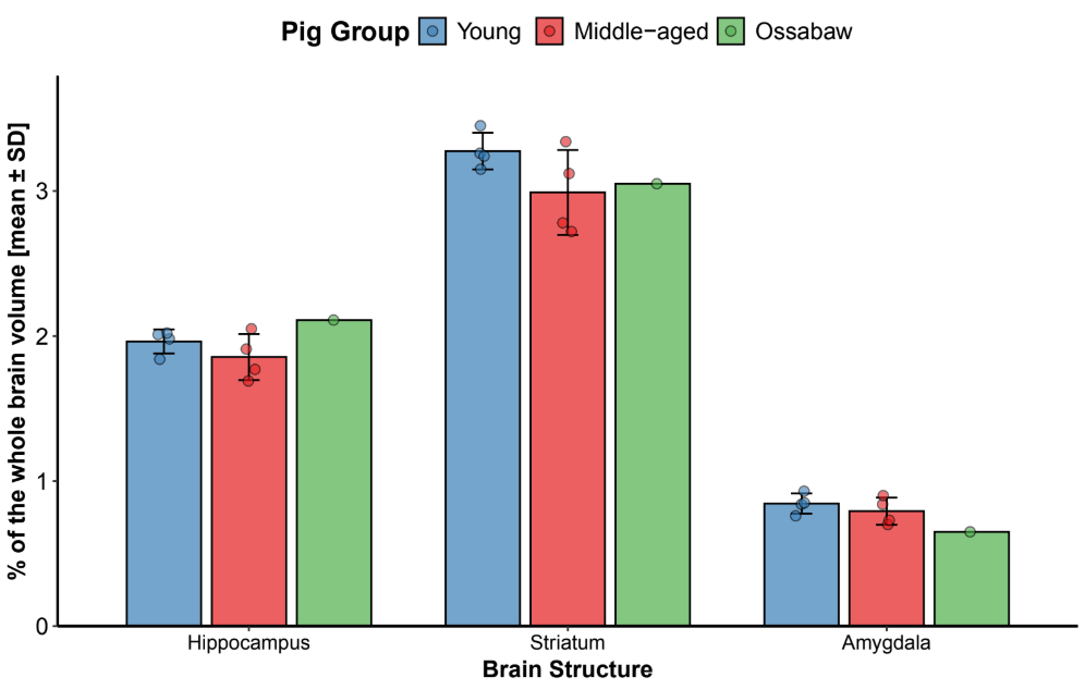
The relative volumes of hippocampus, striatum, and amygdala
(left and right sides summed) in relation to the total brain
volume are shown in Figure 1. The average volume fraction was
slightly lower for the mid-dle-aged minipigs compared to the
young, but the dif-ferences were not significant for either the
hippocam-pus (p=0.30), striatum (p=0.15), or amygdala (p=0.41).
The average volume fractions in the Ossabaw pig were roughly on
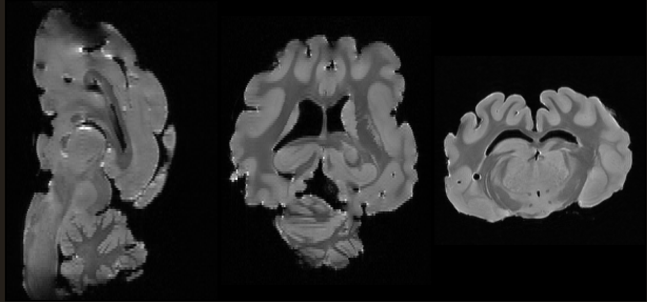
par with what was found in the Göttingen minipigs (Figure 1). MR
images from the oldest (11 years) Göttingen minipig are shown in
Figure 2.
| Pig breed and age group | Age, body weight, and wet brain weight |
Total brain volume (mm3) |
Hippocampus volume (mm3) |
Striatum volume (mm3) |
Amygdala volume (mm3) |
|---|---|---|---|---|---|
| Göttingen minipigs, middle-aged | 4 years; 41.2 kg; 78 g | 80,580 | L: 676 R: 689 Mean: 683 |
L: 1,113 R: 1,123 Mean: 1,118 |
L: 282 R: 281 Mean: 282 |
| 6 years; 47.0 kg; 78 g | 81,310 | L: 711 R: 727 Mean: 719 |
L: 1,250 R: 1,288 Mean: 1,269 |
L: 341 R: 342 Mean: 342 |
|
| 4 years; 41.2 kg; 76.5 g | 79,250 | L: 726 R: 787 Mean: 757 |
L: 1,086 R: 1,066 Mean: 1,076 |
L: 286 R: 291 Mean: 289 |
|
| 11 years; 41.9 kg; 76 g | 73,940 | L: 743 R: 776 Mean: 760 |
L: 1,230 R: 1,240 Mean: 1,235 |
L: 341 R: 327 Mean: 334 |
|
| Göttingen minipigs, young adults | 7 months; 19 kg; wet brain weight unknown | 66,460 | L: 635 R: 679 Mean: 657 |
L: 993 R: 1,099 Mean: 1,046 |
L: 287 R: 272 Mean: 280 |
| 7 months; 17 kg; wet brain weight unknown | 66,695 | L: 615 R: 611 Mean: 613 |
L: 1,076 R: 1,098 Mean: 1,087 |
L: 286 R: 283 Mean: 285 |
|
| 7 months; 16 kg; wet brain weight unknown | 63,983 | L: 647 R: 643 Mean: 646 |
L: 1,099 R: 1,111 Mean: 1,105 |
L: 295 R: 229 Mean: 262 |
|
| 1 year; 18 kg; wet brain weight unknown | 66,820 | L: 660 R: 684 Mean: 672 |
L: 1,133 R: 1,032 Mean: 1,083 |
L: 243 R: 267 Mean: 255 |
|
| Ossabaw, middle-aged | 8 years; 115 kg; 124 g | 121,767 | L: 1,256 R: 1,254 Mean: 1,255 |
L: 1,813 R: 1,821 Mean: 1,817 |
L: 381 R: 387 Mean: 384 |
Note: L, left; R, right
Discussion
The human brain starts to show signs of aging (white matter lesions and atrophy) between the ages of 40 and 50. Aging signs accelerate with age, high blood pres-sure and unhealthy lifestyle factors (Yang et al. 2024). In this study, we compared total brain volumes and selected substructures in young adult and middle-aged Göttingen minipigs, with the additional inclusion of a single Ossabaw pig. If it were possible to make a direct comparison, the age of the examined pig brains would correspond in human years to 40-50 years or younger, but it is unknown how the pig brain deteriorates over time, and this adds to the uncertainty in using the pig brain, when an aged brain is required for scientific re-search. Contrary to expectations, even the oldest min-ipigs examined (up to eleven years of age) showed no clear neuroanatomical signs of aging, such as cortical or subcortical brain atrophy, ventricular enlargement, or region-specific shrinkage, features that are well doc-umented in aging humans and observed in other spe-cies with shorter lifespans (Blinkouskaya et al. 2021). Instead, total brain volumes were larger in middle-aged Göttingen minipigs compared with young adults, and subcortical structures such as the hippocampus, stria-tum, and amygdala were either stable or showed proportional increases. The Ossabaw pig, despite its larger body and brain weight, displayed a similar volumetric pattern. While total brain volume was lower in the 11-year-old than in the 4-year-old Göttingen minipigs; no volume loss was observed in the examined subcor-tical structure
These findings highlight a fundamental limitation in using pigs
as translational models for aging research. Even the “oldest”
laboratory pigs accessible are not physiologically aged in a
neurobiological sense. Pigs have a lifespan of 15–20 years
(Klein 2021), and our middle-aged Göttingen minipigs, aged 4–11
years, may represent middle age rather than true senescence.
This creates a translational gap, as many neurodegenerative
disorders in humans manifest only after midlife (Mas-ters et al.
2015; Boehme et al. 2017). Thus, convention-al laboratory pigs
may not yet exhibit the neurobiologi-cal hallmarks relevant to
late-life neurodegeneration.
Our observations are consistent with reports not-ing that
obtaining aged large animals, including pigs, sheep, and goats,
is difficult (Holtze et al. 2021). This challenge is partly due
to the high costs associated with long-term housing, husbandry
demands, as well as the limited duration of research projects
and grants, which typically do not extend over the decades
required for animals to reach their natural old age.
Consequently, most large animal studies rely on juvenile or
young adult subjects, which may limit translational validity and
potentially introduce bias findings when studying age-related
neurological disorders.
From a translational perspective, pigs/minipigs remain highly
valuable: their gyrencephalic brains, hu-man-like gray/white
matter ratio, and relatively large brain size (contrary to
small, lissencephalic rodent brains) makes them particularly
suitable to be used for neurosurgical and neuromodulation
studies. Likewise, the size of the minipig body allows the use
of standard clinical imaging modalities like CT or MRI (Tohyama
and Kobayashi 2018; Lunney et al. 2021; Sørensen et al. 2011).
The size of the pig brain makes it useful, for instance, for
stroke research (Kuang et al. 2025), deep brain stimulation
studies using human-intended elec-trodes (Orlowski et al. 2017),
or PET studies in e.g., Parkinson's disease models (Lillethorup
et al. 2018). For aging studies, the pig may also be useful. Age
is a leading predictor of disease (An et al. 2022),
con-sequently, understanding which biological parameters change
with age and how these factors evolve is cru-cial. Animal model
validity is, therefore, a main concern in the study of the aging
brain. The human brain is estimated to lose roughly 5% of its
volume per dec-ade after the age of 40 (Markov et al. 2022).
Based on observations, veterinarians have established
correlation tables to convert pig age to human age equivalents
by multiplying the pig’s age in years by five to estimate its
human age equivalent
(https://www.minipiginfo.com/estimating-the-age-of-a-mini-pig.html).
Based on this, the oldest pig included in this study (11 years
of age) would correspond approximately to a human aged 50-60
years. This is, of course, a rough estimate as pig age
equivalence may be more reliable for young pigs, as pigs grow
rapidly and mature early (Tohyama and Kobayashi 2018). If,
however, one assumes similar ag-ing patterns in the human and
pig brain it is therefore surprising that more atrophy is not
seen in the rather middle-aged pigs investigated here. Recent
studies in-dicate that the aging process in the brain is
accelerated by chronic, high levels of proinflammatory immune
factors (Markov et al. 2022; Tamatta et al. 2025). It may
therefore be that the controlled environments that laboratory
pigs live in shield them from some of the factors that drive
aging-related atrophy in the human brain. Usually, the
well-controlled conditions of animal studies are considered an
advantage in the effort to isolate disease effects, but here it
would seem that the lack of environmental factors should be
considered in animal-based studies aiming to mimic normal human
brain aging. If so, this is likely the case for most labo-ratory
animal species, underscoring the need to study brain aging in
humans alongside studies in animal mod-els.
From a methodological perspective, the design of our study was
suboptimal and demonstrated addition-al complications. Brains
were obtained post-mortem from different sources, with variable
fixation methods, and scanned after prolonged immersion
fixation. Such inconsistencies may contribute to volume
distortions, as formalin fixation is known to cause tissue
shrink-age (de Guzman et al. 2016). Furthermore, our small
sample size, particularly the single Ossabaw pig, lim-its
statistical generalization. However, we conducted the study
based on a 3R approach to reduce the use of animals, as no
animals were euthanized specifical-ly for this study. Even
though we collected the oldest research pig brains available,
they still showed no signs of aging. Nevertheless, the
consistency of the findings across Göttingen minipigs suggests
that the lack of at-rophy is not an artifact caused by these
limitations but rather reflects the biology of pigs at the ages
we were able to obtain.
Another critical issue is the species-specific tra-jectory of
aging. Aging is a complex, multifactorial process (Cohen 2018).
Rodents show pronounced and relatively early onset of brain
aging (Radulescu et al. 2021), allowing convenient modeling of
human neuro-degeneration, even if their brain is non-gyrated.
Gyrated brains from larger mammals such as pigs may age more
slowly, with structural and metabolic changes manifesting later
in life (Cohen 2018). Thus, pigs may still serve as valuable
models of other aspects of hu-man neurobiology, such as the
study of brain structure, connectivity, physiology, or response
to various factors, as well as symptomatic modeling of the
various diseas-es (Bjarkam et al. 2017; Orlowski et al. 2017;
Bech et al. 2018; Lillethorup et al. 2018; Orstrup et al. 2019;
Win-terdahl et al. 2019; Bech et al. 2020; Zaer et al. 2020;
Zaer et al. 2022; Kuang et al. 2025), but their utility in aging
research is limited unless access to old animals becomes easier.
Moreover, studies of aging should be carefully planned and
adjusted for every research ques-tion, including the choice of
experimental animal models.
The future solution of the problems associated with studying
aging using large animal models may include developing
collaborative repositories of aged large animal tissues,
integrating naturally aged farm or companion animals into
research, or applying acceler-ated-aging interventions (e.g.,
genetic or metabolic ma-nipulations) (Azman and Zakaria 2019;
Liu et al. 2020; Cai et al. 2022) in pigs to approximate human
late-life neurobiology. Alternatively, studies may need to rely
on cross-species comparative approaches, integrating ro-dent
models for aging with pig or non-human primate models for
anatomy and network-level analyses. An-other approach could
involve large-scale epidemiolog-ical studies in human
populations, such as NHANES (Crimmins et al. 2008), and
well-established longitu-dinal cohorts like ELSA (The English
Longitudinal Study of Ageing), DanACo (The Danish Aging and
Cognition) and SHARE (The Survey of Health, Age-ing and
Retirement in Europe) (Wigmore et al. 2017; Ruiz-Adame et al.
2023; Gronkjaer et al. 2024; Rosenau et al. 2024), combining
biomarker identification (Moqri et al. 2024) across multiple
physiological domains with population-level and correlational
studies of age-related diseases.
To conclude, although pigs hold promise as trans-lational models
for neuroscience, our findings indicate that currently
accessible middle-aged animals do not exhibit the anatomical
hallmarks of brain aging. Even at 11 years of age, Göttingen
minipigs lack the struc-tural signatures of neurodegeneration.
Thus, critical aspects of late-life neurodegeneration cannot be
cap-tured in available pig models unless studies can access
substantially older animals or novel aging-acceleration
approaches are adopted.
Acknowledgements
No financial funding was obtained for this study. The authors would like to thank the animal facility at the Foulum Research Center, Aarhus University, as well as the DTU European Ossabaw facility for donating the brains for this study. Thanks to Michele Gammeltoft for linguistic proofreading
Conflict of interests
The authors declare that they have no conflicts of interest.
References
- An, S., Ahn, C., Moon, S., Sim, E.J., Park, S.-K., (2022). Individualized biological age as a predictor of disease: Korean genome and epidemiology study (KoGES) cohort. Journal of Personalized Medicine. 12(3), 555. doi: 10.3390/jpm12030505
- Ascherio, A., Schwarzshild, M.A., (2016). The epidemiology of Parkinson´s disease: risk factors and prevention. The Lancet Neurology. 15(12), 1257-1272. doi: 10.1016/S1474-4422(16)30230-7.
- Azman, K.F., Zakaria, R., (2019). D-Galactose-induced accelerated aging model: an overview. Biogerontology. 20(6), 763-782. doi: 10.1007/s10522-019-09837-y.
- Bartolomucci, A., Kane, A.E., Gaydosh, L., Razzoli, M., McCoy, B.M., Ehninger, D., Chen, B.H., Howlett, S.E., Snyder-Mackler, N., (2024). Animal models relevant for geroscience: Current trends and future perspectives in biomarkers, and measures of biological aging. Journal of Gerontology A - Biological Sciences and Medical Sciences. 79(9), 135. doi: 10.1093/gerona/glae135.
- Bech, J., Glud, A.N., Sangill, R., Petersen, M., Frandsen, J., Orlowski, D., West, M.J., Pedersen, M., Sorensen, J.C.H., Dyrby, T.B., Bjarkam, C.R., (2018). The porcine corticospinal decussation: A combined neuronal tracing and tractography study. Brain Research Bulleting. 142, 253-262. doi: 10.1016/j.brainresbull.2018.08.004.
- Bech, J., Orlowski, D., Glud, A.N., Dyrby, D.B., Sorensen, J.C.H., Bjarkam, C.R., (2020). Ex vivo diffusion-weighted MRI tractography of the Gottingen minipig limbic system. Brain Structure and Functions. 225(3), 1055-1071. doi: 10.1007/s00429-020-02058-x.
- Bjarkam, C.R., Glud, A.N, Orlowski, D., Sorensen, J.C.H., Palomero-Gallagher, N., (2017). The telencephalon of the Gottingen minipig, cytoarchitecture and cortical surface anatomy. Brain Structure and Functions. 222(5), 2093-2114. doi: 10.1007/s00429-016-1327-5.
- Blinkouskaya, Y., Cacoilo, A., Gollamudi, T., Jalalian, S., Weickenmeier, J., (2021). Brain aging mechanisms with mechanical manifestations. Mechanisms of Ageing and Development. 200, 111575. doi: 10.1016/j.mad.2021.111575.
- Boehme, A.K., Esenwa, C., Elkind, M.S.V., (2017). Stroke risk factors, genetics, and prevention. Circulation Research. 120(3), 472-495. doi: 10.1161/CIRCRESAHA.116.308398.
- Cai, N., Wu, Y., Huang, Y., (2022). Induction of accelerated aging in a mouse model. Cells. 11(9),1418. doi: 10.3390/cells11091418.
- Cohen, A.A., (2018). Aging across the tree of life: The importance of a comparative perspective for the use of animal models in aging. Biochimica et Biophysica Acta Molecular Basis of Diseases. 1864(9 Pt A), 2680-2689. doi: 10.1016/j.bbadis.2017.05.028.
- Crimmins, E., Vasunilashorn, S., Kim, J.K., Alley, D., (2008). Biomarkers related to aging in human populations. Advances in Clinical Chemistry. 46, 161-216. doi: 10.1016/s0065-2423(08)00405-8.
- de Guzman, A.E., Wong, M.D., Gleave, J.A., Nieman, B.J., (2016). Variations in post-perfusion immersion fixation and storage alter MRI measurements of mouse brain morphometry. Neuroimage. 142, 687-695. doi: 10.1016/j.neuroimage.2016.06.028.
- Dohm-Hansen, S., English, J.A., Lavelle, A., Fitzsimons, C.P., Lucassen, P.J., Nolan, Y.M., (2024). The 'middle-aging' brain. Trends in Neuroscience. 47(4):259-272. doi:10.1016/j.tins.2024.02.001.
- Félix, B., Léger, M.E., Albe-Fessard, D., Marcilloux, J.C., Rampin, O., Laplace, J.P., (1999). Stereotaxic atlas of the pig brain. Brain Research Bulletin. 49(1-2), 1-137. doi: 10.1016/s0361-9230(99)00012-x. PMID: 10466025.
- Gronkjaer, M., Mortensen, E.L., Wimmelmann, C.L., Flensborg-Madsen, T., Osler, M., Okholm, G.T., (2024). The Danish aging and cognition (DanACo) cohort. BMC Geriatrics. 24(1), 238. doi: 10.1186/s12877-024-04841-5.
- Holtze, S., Gorshkova, E., Braude, S., Cellerino, A., Dammann, P., Hildebrandt, T.B., Hoeflich, A., Hoffmann, S., Koch, P., Terzibasi Tozzini, E., Skulachev, M., Skulachev, V.P., Sahm, A., (2021). Alternative animal models of aging research. Frontiers in Molecular Bioscience. 8, 660959. doi: 10.3389/fmolb.2021.660959.
- Kim, S., Kwon, S.-H., Kam, T.-I., Panicker, N., Karuppagounder, S.S., Lee, S., Lee, J.H., Kim, W.R., Kook, M., Foss, C.A., Shen, C., Lee, H., Kulkarni, S., Pasricha, P.J., Lee, G., Pomper, M.G., Dawson, V.L., Dawson, T.M., Ko, H.S., (2019). Transneuronal propagation of pathologic alpha-synuclein from the gut to the brain models Parkinson´s disease. Neuron. 103(4), 627-641. doi: 10.1016/j.neuron.2019.05.035
- Klein, V., (eds.) (2019). Cummingham´s textbook of veterinary physiology. 6th. Ed.: Saunders. DOI: 10.1016/C2015-0-06149-4.
- Klæstrup, I.H., Just, M.K., Holm, K.L., Alstrup, A.K.O., Romero-Ramos, M., Borghammer, P., Van Den Berge, N., (2022). Impact of aging on animal models of Parkinson´s disease. Frontiers in Aging Neuroscience. 14, 909273. doi: 10.3389/fnagi.2022.909273.
- Knopper, R.W., Skoven, C.S., Eskildsen, S.F., Østergaard, L., Hansen, B., (2024). The effects of locus coeruleus ablation on mouse brain volume and microstructure evaluated by high-field MRI. Frontiers in Cellular Neuroscience. 18, 1498133. doi: 10.3389/fncel.2024.1498133.
- Kuang, V.H., Skoven, C.S., Arvin, S., Fitting, L.M., Drasbek, K.R., Hansen, B., Orlowski, D., Sorensen, J.C.H., (2025). A large animal model for focal stroke: Photothrombotic lesion in the cortex of Danish Landrace pigs. Journal of Neuroscience Methods. 418, 110408. doi: 10.1016/j.jneumeth.2025.110408.
- Lee, J., Kim, H.J., (2022). Normal aging induces changes in the brain and neurodegeneration progress: Review of the structural, biochemical, metabolic, cellular, and molecular changes. Frontiers in Aging Neuroscience. 14, 931536. doi:10.3389/fnagi.2022.931536.
- Lillethorup, T.P., Glud, A.G., Alstrup, A.K.O., Noer, O., Nielsen, E.H.T., Schacht, A.C., Landeck, N., Kirik, D., Orlowski, D., Sorensen, J.C.H., Doudet, D.J., Landau, A.M., (2018). Longitudinal monoaminergic PET imaging of chronic proteasome inhibition in minipigs. Scientific Reports. 8(1), 15715.
- Liu, B., Liu, J., Shi, J.S., (2020). SAMP8 mice as a model of age-related cognition decline with underlying mechanisms in Alzheimer's disease. Journal of Alzheimers Disease. 75(2), 385-395. doi: 10.3233/JAD-200063.
- Lunney, J.K., Goor, A., Walker, K.E., Hailstock, T., Franklin, J., Dai, C., (2021). Importance of the pig as a human biomedical model. Science Translational Medicine. 13, 621. doi: 10.1126/scitranslmed.abd5758.
- Markov, N.T., Lindbergh, C.A., Staffaroni, A.M., Furman, D., (2022). Age-related brain atrophy is not a homogenous process: Different functional brain networks associate differentially with aging and blood factors. The Proceedings of the National Academy of Sciences. 119(49): e2207181119. doi: 10.1073/pnas.2207181119.
- Masters, C.L., Bateman, R., Blennow, K., Rowe, C.C., Sperling, R.A., Cummings, J.L., (2015). Alzheimer´s disease. Nature Reviews Disease Primers. 1, 15056. doi: 10.1038/nrdp.2015.56.
- Moqri, M., Herzog, C., Poganik, J.R., Ying, K., Justice, J.N., Belsky, D.W., Higgins-Chen, A.T., Chen, B.H., Cohen, A.A., Fuellen, G., Hagg, S., Marioni, R.E., Widschwendter, M., Fortney, K., Fedichev, P.O., Zhavoronkov, A., Barzilai, N., Lasky-Su, J., Kiel, D.P., Kennedy, B.K., Cummings, S., Slagboom, P.E., Verdin, E., Maier, A.B., Sebastiano, V., Snyder, M.P., Gladyshev, V.N., Horvath, S. Ferrucci, L., (2024). Validation of biomarkers of aging. Nature Medicine. 30(2), 360-372. doi: 10.1038/s41591-023-02784-9.
- Orlowski, D., Michalis, A., Glud, A.N., Korshoj, A.R., Fitting, L.M., Mikkelsen, T.W., Mercanzini, A., Jordan, A., Dransart A., Sorensen, J.C.H., (2017). Brain tissue reaction to deep brain stimulation - A longitudinal study of DBS in the Goettingen minipig. Neuromodulation. 20(5), 417-423. doi: 10.1111/ner.12576.
- Orlowski, D., Glud, A.N., Palomero-Gallagher, N., Sørensen, J.C.H., Bjarkam, C.R., (2019). Online histological atlas of the Göttingen minipig brain. Heliyon. 5(3), e01363. doi: 10.1016/j.heliyon.2019.e01363.
- Orstrup, L., Tvilling, H.L., Orlowski, D., Zaer, H., Bjarkam, J.R., von Voss, P., Andersen, P.S., Christoffersen, B.O., Hedemann Sorensen, J.C, Laursen, T., Thygesen, P., Lykkesfeldt J., Glud, A.N., (2019). Towards a Gottingen minipig model of adult onset growth hormone deficiency: evaluation of stereotactic electrocoagulation method. Heliyon. 5(11), e02892. doi: 10.1016/j.heliyon.2019.e02892.
- Padmanabhan, P., Gotz, J., (2023). Clinical relevance of animal models in aging-related dementia research. Nature Aging. 3(5), 481-493. doi: 10.1038/s43587-023-00402-4. Radulescu, C.I., Cerar, V., Haslehurst, P., Kopanitsa, M., Barnes, S.J., (2021). The aging mouse brain: cognition, connectivity and calcium. Cell Calcium. 94: 102358. doi: 10.1016/j.ceca.2021.102358.
- Rosenau, C., Kohler, S., van Boxtel, M., Tange, H., Deckers, K., (2024). Validation of the updated "LIfestyle for BRAin health" (LIBRA) index in the English longitudinal study of ageing and Maastricht aging study. Journal of Alzheimers Disease. 101(4), 1237-1248. doi: 10.3233/JAD-240666.
- Ruiz-Adame, M., Ibanez, A., Mollayeva T., Trepel D., (2023). Association between neuroticism and dementia on healthcare use: A multi-level analysis across 27 countries from the survey of health, ageing and retirement in Europe (SHARE). Journal of Alzheimers Disease. 95(1), 181-193. doi: 10.3233/JAD-230265.
- Saikali, S., Meurice, P., Sauleau, P., Eliat, P.A., Bellaud, P., Randuineau, G., Vérin, M., Malbert, C.H., (2010). A three-dimensional digital segmented and deformable brain atlas of the domestic pig. Journal of Neuroscience Methods. 192(1), 102-9. doi: 10.1016/j.jneumeth.2010.07.041.
- Shepherd, T.M., Thelwall, P.E., Stanisz, G.J., Blackband, S.J., (2009). Aldehyde fixative solutions alter water relaxation and diffusion properties of nervous tissue. Magnetic Resonance in Medicine. 62(1), 26-34. doi: 10.1002/mrm.21977.
- Sørensen, J.C., Nielsen, M.S., Rosendal, F., Deding, D., Ettrup, K.S., Jensen, K.N., Jørgensen, R.L., Glud, A.N., Meier, K., Fitting, L.M., Møller, A., Alstrup, A.K.O., Ostergaard, L., Bjarkam, C.R., (2011). Development of neuromodulation treatments in a large animal model--do neurosurgeons dream of electric pigs? Progress in Brain Research. 194, 97-103. doi: 10.1016/B978-0-444-53815-4.00014-5.
- Suter, P., Luetkemeier, H., Zakova, N., Christen, P., Sachsse K., Hess, R., (1979). Lifespan studies on male and female mice and rats under SPF-laboratory conditions. Archives of Toxicology. 2, 403-407.
- Tamatta, R., Pai, V., Jaiswal, C., Singh, I., Singh, A.K., (2025). Neuroinflammaging and the immune landscape: The role of autophagy and senescence in aging brain. Biogerontology. 26(2), 52. doi: 10.1007/s10522-025-10199-x.
- Tohyama, S., Kobayashi, E., (2018). Age-appropriateness of porcine models used for cell transplantation. Cell Transplantation. 28(2), 2024-228. doi: 10.1177/0963689718817477.
- Tustison, N.J., Avants, B.B., Cook, P.A., Zheng, Y., Egan, A., Yushvich, P.A., Gee, J.C., (2010). N4ITK: improved N3 bias correction. IEEE Transaction on Medical Imaging. 29(6): 1310-1320. doi: 10.1109/TMI.2010.2046908.
- Van Den Berge, N., Ferreira, N., Gram, H., Mikkelsen, T.W., Alstrup, A.K.O., Casadei, N., Tsung-Pin, P., Riess, O., Nyengaard, J.R., Tamgüney, G., Jensen, P.H., Borghammer, P., (2019). Evidence for bidirectional and trans-synaptic parasympathetic and sympathetic propagation of alpha-synuclein in rats. Acta Neuropatology. 138(4), 535-550. doi: 10.1007/s00401-019-02040-w.
- Van Den Berge, N., Ferreira, N., Mikkelsen, T.W., Alstrup, A.K.O., Tamgüney, G., Karlsson, P., Terkelsen, A.J., Nyengaard, J.R., Jensen P.H., Borghammer, P., (2021). Ageing promotes pathological alpha-synucleinpropagation and autonomic dysfunction in wild-type rats. Brain. 133(6), 1853-1868. doi: 10.1093/brain/awab061.
- Veraart, J., Novikov, D.S., Christians, D., Ades-Aron, B., Sijbes, J., Fieremans, E., (2016). Denoising of diffusion MRI using random matrix theory. Neuroimage. 142, 394-406. doi: 10.1016/j.neuroimage.2016.08.016.
- Wigmore, E.M., Clarke, T.K., Howard, D.M., Adams, M.J., Hall, L.S., Zeng, Y., Gibson, J., Davies, G., Fernandez-Pujals, A.M., Thomson, P.A., Hayward, C., Smith, B.H., Hocking, L.J., Padmanabhan, S., Deary, I.J., Porteous, D.J., Nicodemus, K.K., McIntosh, A.M., (2017). Do regional brain volumes and major depressive disorder share genetic architecture? A study of generation Scotland (n=19 762), UK Biobank (n=24 048) and the English longitudinal study of ageing (n=5766). Translational Psychiatry 7(8), e1205. doi: 10.1038/tp.2017.148.
- Winterdahl, M., Noer, O., Orlowski, D., Schacht, A.C., Jakobsen, S., Alstrup, A.K.O., Gjedde, A., Landau, A.M., (2019). Sucrose intake lowers mu-opioid and dopamine D2/3 receptor availability in porcine brain. Scientific Reports. 9(1), 16918. doi: 10.1038/s41598-019-53430-9.
- Wood, H., (2019). New models show gut-brain transmission of Parkinson disease pathology. Nature Reviews Neurology. 15: 491. doi: 10.1038/s41582-019-0241-x.
- Yushkevich, P.A., Piven, J., Hazlett, H.C., Smith, R.G., Ho, S., Gee, J.C., Gerig, G., (2006). User-guided 3D active contour segmentation of anatomical structures: significantly improved efficiency and reliability. Neuroimage. 31(3), 1116-1128. doi: 10.1016/j.neuroimage.2006.01.015.
- Yang, Z., Wen, J., Erus, G., Govindarajan, S.T., Melhem, R., Mamourian, E., Cui, Y., Srinivasan, D., Abdulkadir, A., Parmpi , P., Wittfeld, K., Grabe, H.J., Bülow, R., Frenzel, S., Tosun, D., Bilgel, M., An, Y., Yi, D., Marcus, D., LaMontagne, P., Benzinger, T.L.S., Heckbert, S.R., Austin, T.R., Waldstein, S.R., Evans, M.K., Zonderman, A.B., Launer, L.J., Sotiras, A., Espeland, M.A., Masters, C.L., Maruff, P., Fripp, J., Toga, A.W., O'Bryant, S., Chakravarty, M.M., Villeneuve, S., Johnson, S.C., Morris, J.C., Albert, M.S., Yaffe, K., Völzke, H., Ferrucci, L., Bryan, R.N., Shinohara, R.T., Fan, Y., Habes, M., Lalousis, P.A., Koutsouleris, N., Wolk D.A , Resnick, S.M., Shou, H., Nasrallah, I., Davatzikos, C., (2024). Brain aging patterns in a large and diverse cohort of 49,482 individuals. Nature Medicine. 30(10), 3015-3026. doi: 10.1038/s41591-024-03144-x.
- Zaer, H., Fan, W., Orlowski, D., Glud, A.N., Jensen, M.B., Worm, E.S., Lukacova, S., Mikkelsen, T.W., Fitting, L.M., Jacobsen, L.M., Portmann, T., Hsieh, J.Y., Noel, C., Weidlich, G., Chung, W., Riley, P., Jenkins, C., Adler Jr, J.R, Schneider, M.B., Sorensen J.C.H., Stroh, A., (2022). Non-ablative doses of focal ionizing radiation alters function of central neural circuits. Brain Stimulation. 15(3), 586-597. doi: 10.1016/j.brs.2022.04.001. doi: 10.1016/j.brs.2022.04.001.
- Zaer, H., Glud, A.N., Schneider, B.M., Lukacova, S., Hansen, K.V., Adler, J.R., Hoyer, M., Jensen, M.B., Hansen, R., Hoffmann, L., Worm, E.S., Sorensen, J.C.H., Orlowski, D., (2020). Radionecrosis and cellular changes in small volume stereotactic brain radiosurgery in a porcine model. Scientific Reports. 10(1), 16223. doi: 10.1038/s41598-020-72876-w.