Joonistused ja fotod mikroskoobipreparaatidest Tartu Ülikooli muuseumi kogudes
Photos and drawings of microscopic preparations in the museum of the University of Tartu
DOI:
https://doi.org/10.15157/tyak.v51i.23356Abstract
Microscopes are fascinating tools that can make the invisible visible.
They are useful educational and scientific aids that have been used
for a few hundred years and besides real life observations, photos and
drawings of what can be seen have been captured. The museum of the
University of Tartu has a collection of various drawings and photos of
microscopic preparations that have been used in teaching medicine
at the university.
These photos and drawings have significant historic, scientific
and cultural value. Many photos in this collection have their author
listed as Dr Kull, whereas most drawings do not have an author listed.
Drawings have captions in Estonian as well as some in Russian;
however, originally, these were written in German using Latin terminology.
It is assumed that most of the drawings were likely illustrated
by university students when making preparations and using
microscopes as part of their practicum. It is assumed that these, now
useful study aids and fascinating museum items, were made in the
first half of the 20th century.
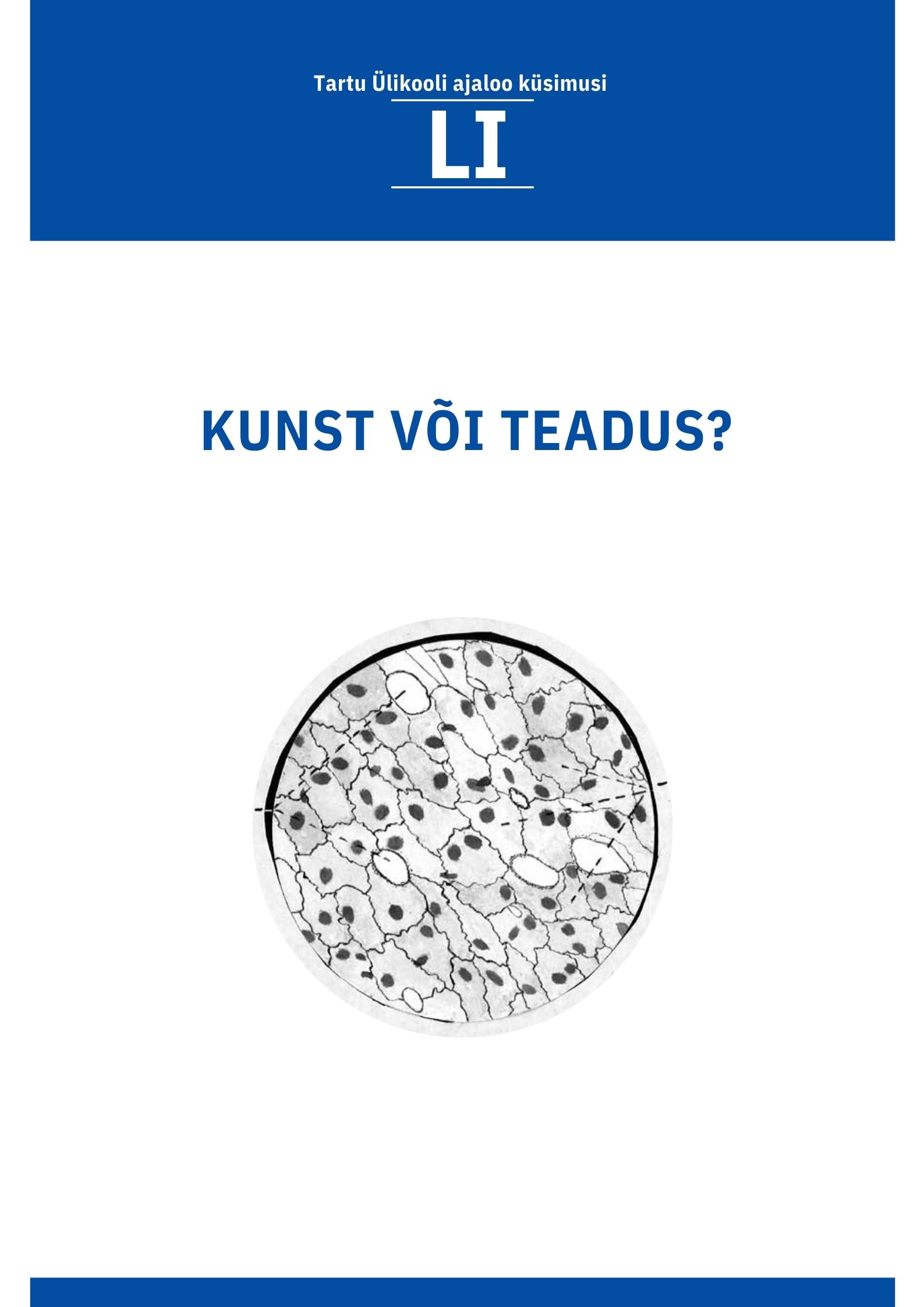
The collection of specimens used in these photos and drawings
is diverse. The majority of them are of various human and animal
tissues, along with some photos and drawings which are able to show
processes such as cell proliferation. Even the processes happening in
the nucleus of a cell can be seen. Microscopic views of human bone,
pancreas, ovary, kidney and even an unborn child’s eye are all examples
of what can be seen in these interesting photos and drawings.
Downloads